40 labeled histology slides
Anatomy Histology Slide for Mbbs Student 1st Year - Part 2 ANATOMY HISTOLOGY SLIDE FOR MBBS STUDENT 1ST YEAR - PART 2 Sahil Alam September 04, 2022 . ANATOMY HISTOLOGY SLIDE FOR MBBS STUDENT 1ST YEAR - PART 2. HISTOLOGY SLIDE RAPID REVISION FOR MBBS FIRST YEAR. HISTOLOGY SLIDE PART 3. Newer. Older. Post a Comment. 0 Comments. AD Ad Space Popular Posts New: Human Anatomy Atlas 2023+, Where 3D Visualization Meets 2D Realism! Welcome to Human Anatomy Atlas 2023+!Now, more than 6,000 manipulatable 3D structures and more than 200 histology slides and illustrations are at your fingertips. The team at Visible Body is excited to launch the newest version of our award-winning Human Anatomy Atlas app. Human Anatomy Atlas 2023+ is the first version to include 2D illustrations and histology slides in addition to our ...
Online Atlases, Histology Images & Study Guides - Anatomy and ... Histology Guide teaches the visual art of recognizing the structure of human cells and tissues and understanding how it is determined by their function. This site contains more than 225 virtual microscope slides and a 100 electron micrographs for the learning human histology.

Labeled histology slides
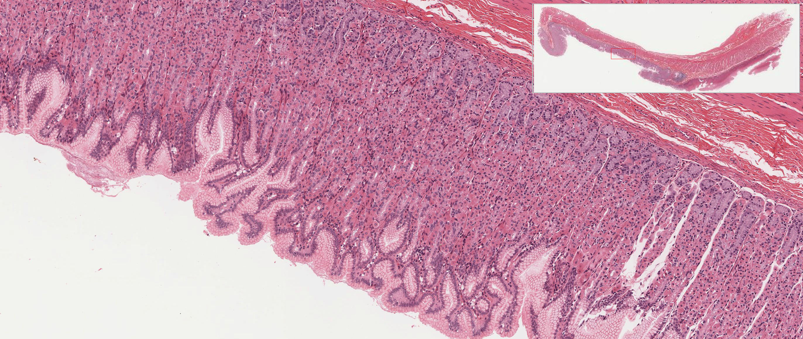
Colon Histology Slide with Labeled Diagram - AnatomyLearner Colon Histology Slide with Labeled Diagram 04/06/2022 04/06/2022 by anatomylearner The colon histology slide possesses the typical four layers of a tubular organ - mucosa, submucosa, muscularis, and serosa. But, there are no permanent plica circularis and villi in the colon slide as found in the different segments of the small intestine. Histology - An Essential Textbook PDF Free Download Learn to identify histological structures and their correlated functions! Histology: An Essential Textbook is a concise, multimedia study guide for medical students who need to learn the functions and related correlations of cells, tissues, and organs of the human body. Professor D.J. Lowrie, Jr. has written a unique and practical medical ... Anatomy, Histology, & Pathology | Continuing Studies Investigate the appearance of cells in various organ systems, including cardiovascular, digestive, respiratory, reproductive and nervous. After seeing the normal tissue state, you will explore the appearance of cells in various disease states (pathology), such as bone osteoporosis, myocardial heart infarction, neoplasms and more.
Labeled histology slides. Spleen histology: Location, functions, structure | Kenhub Spleen histology slide (labeled) The spleen is a fist sized organ located in the left upper quadrant of the abdomen. It is the largest lymphoid organ and thus the largest filter of blood in the human body. The spleen has a unique location, embryological development and histological structure that differs significantly from other lymphoid organs. Lymph nodes: Histology | Kenhub Lymph node (histological slide) The presence of foreign organisms within the blood stream can trigger a massive cascade of events that will disrupt many homeostatic microenvironments within the body. Therefore, the immune system carries out detailed surveillance of the blood in order to detect these pathogens. Histology, Lung - StatPearls - NCBI Bookshelf The lung is identified, dissected en-block, weighed, and labeled. Later the lung is perfused with 10% formalin through the trachea to the physiological peak inspiration level. ... (IHC). Both methods can be performed on tissue slides. The normal lung cells can be typed as type I and type II pneumocytes based on histochemical ... By histology ... Learn histology faster With quizzes and flashcards | Kenhub With Kenhub's huge library of histology slides, of course! In our histology atlas, we clearly highlight a given structure on our slides. Comparing several of these slides next to one another is a great way to get a feel for how one tissue differs from another. Enter: our labeled and unlabeled histology tissue identification quiz worksheets.
Jejunum Histology Slide with Labeled Diagram and ... - AnatomyLearner Jejunum Histology Slide with Labeled Diagram and Identification Points 29/10/2021 29/10/2021 by anatomylearner The jejunum is the second part of the small intestine of an animal. In the jejunum histology, you will find the four different layers like a tubular organ with some other specific identifying features. Fallopian tube histology slide diagram and identification points Fallopian tube histology slide diagram The mucosal fold is lined by simple ciliated columnar epithelium You will find nonciliated peg (secretory) cells among the ciliated columnar cells with still higher magnification. Presence of smooth muscle fibers that are arranged in two layers, inner circular and outer longitudinal. Netter's Essential Histology PDF 3rd Edition free Download: Netter's Essential Histology, 3rd Edition, is the perfect text for today's evolving medical education. Concise and easy to use, it integrates gross anatomy and embryology with classic histology slides and state-of-the-art scanning electron microscopy, offering a clear, visual understanding of this complex subject. Histology, Staining - StatPearls - NCBI Bookshelf Medical Histology is the microscopic study of tissues and organs through sectioning, staining, and examining those sections under a microscope. Often called microscopic anatomy and histochemistry, histology allows for the visualization of tissue structure and characteristic changes the tissue may have undergone. Because of this, it is utilized in medical diagnosis, scientific study, autopsy ...
Testes Slide Labeled - lab microscope slide list flashcards easy ... Testes Slide Labeled. Here are a number of highest rated Testes Slide Labeled pictures on internet. We identified it from honorable source. Its submitted by presidency in the best field. We receive this nice of Testes Slide Labeled graphic could possibly be the most trending topic later than we ration it in google improvement or facebook. Slides - HistologySlide by histologyslide Adrenal gland histology slide diagram and identification points The adrenal gland consists of a yellow peripheral region (the cortex), and a brown central region, the medulla. Here, you will learn all the features from the adrenal gland histology slide with a labeled diagram and perfect identification points. Ovary histology slide diagram and identification points In the ovary histology slide labeled diagram, you will find all the features that are mentioned above. Identification of other slides from the female organs Uterine tube histology slide with its identification points Uterus histology slide with its identification points Mammary glands histology slide with its identification points Anatomy Histology Slide for Mbbs Student 1st Year - Part 2 anatomy histology slide for mbbs student 1st year - part 2 0 sahil alam september 04, 2022 . anatomy histology slide for mbbs student 1st year - part 2. histology slide rapid revision for mbbs first year. histology slide part 3. tags. histology.
Histology Virtual Lab - Epithelial Tissues
Rumen Histology Slide Identification with Labeled Diagram Omasum histology slide labeled The tunica submucosa of omasum is comparatively thinner than that of rumen or reticulum. You will find thin outer longitudinal and thicker inner circular layers of smooth muscle in the tunica muscularis of the omasum. The innermost fibers of the circular layer are continued into the larger omasal laminae.
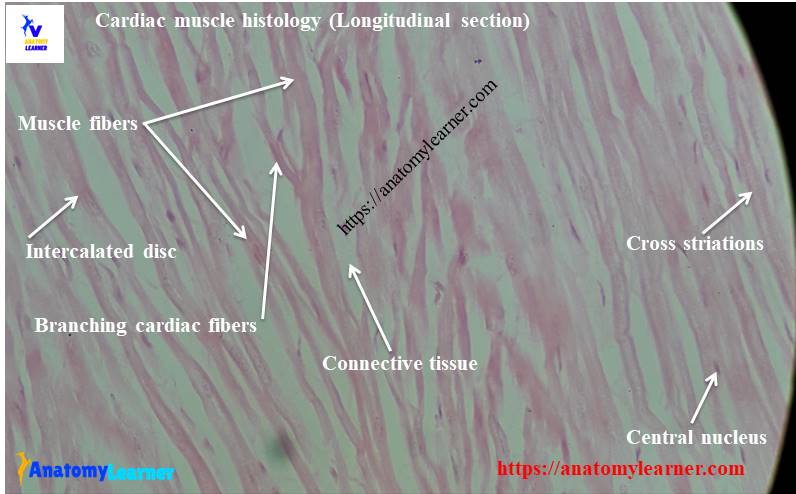
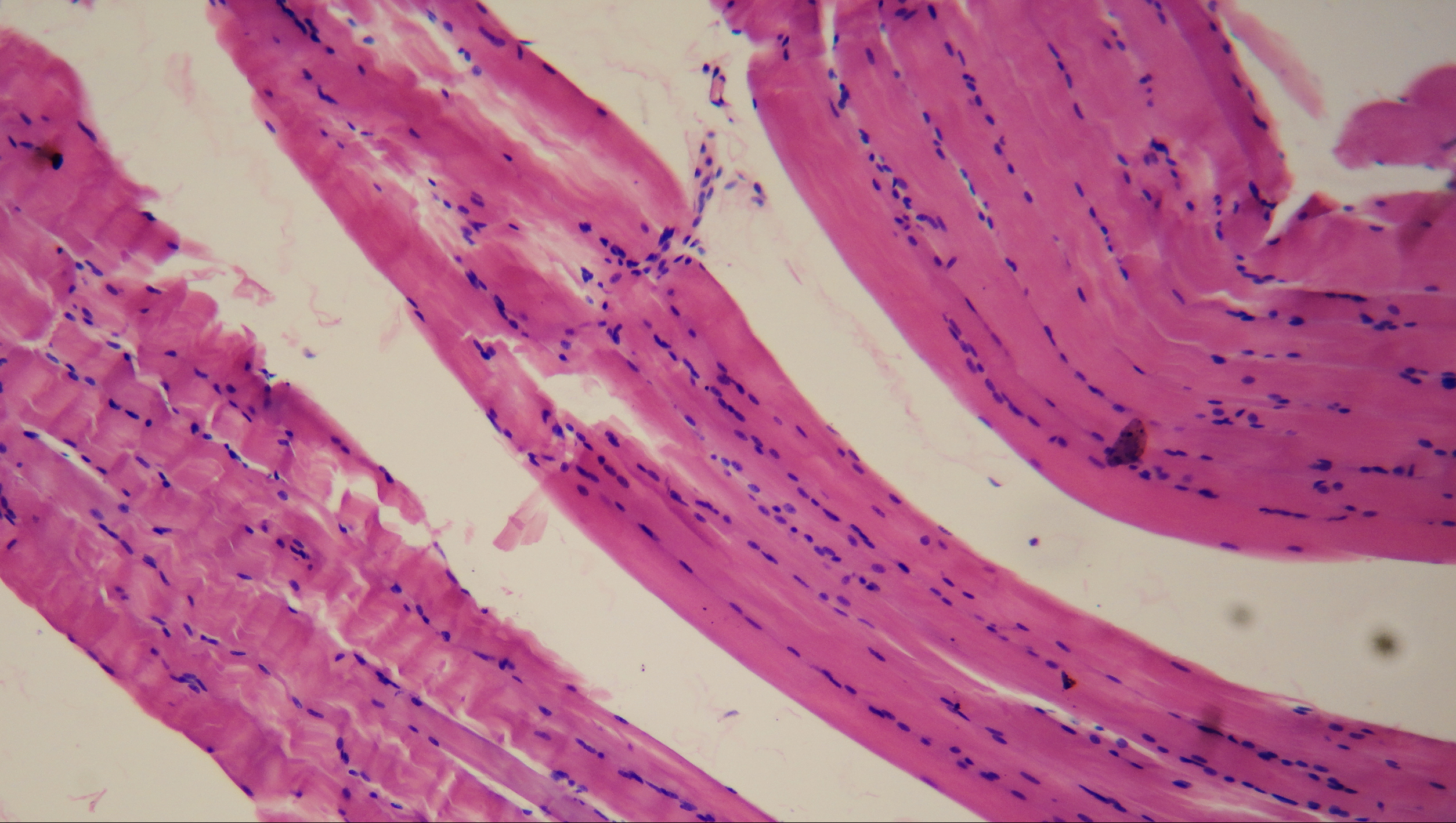
Cardiac Muscle Histology and Slide Identification Points ...
Cecum Histology Slide with Labeled Image and Diagram Generally, the tunica muscularis layer of the cecum histology slide consists of a thick inner circular and thin outer longitudinal smooth muscle layer. The outer longitudinal smooth muscle layer forms large, flat, smooth muscle bands in pig and horse cecum. This smooth muscle band of the pig and horse cecum is known as the taenia Ceci.
Revision of histology slides
Anatomy Resources: Histology Resources - George Washington University Histology Resources. This resources, from Virginia Commonwealth University, is an open educational resource that combines a digital atlas with extensive descriptive text. It is organized as a multi-hierarchy outline that reinforces broader histological concepts and parallels the content of most histology textbooks. Features on-demand labeling ...
Histology Slides 1
Blood Histology Slides with Description and Labeled Diagram In the blood histology slide (smear), all of the cells mentioned earlier with their useful identifying features. There are five different types of leukocytes in blood - neutrophils, eosinophils, basophils, monocytes, and lymphocytes. The neutrophils, eosinophils, and basophils are known as the polymorphonuclear granulocytes.
Solved] Please label the histology slides for urinary system ...
Histology guide: Definition and slides | Kenhub At a histological level, both the heart and blood vessels consist of three layers: Endothelial layer - epithelial tissue formed by simple squamous (endothelial) cells. In the heart, this layer is referred to as endocardium. Muscular layer - smooth muscle in the blood vessels, cardiac muscle (myocardium) in the heart.
File:Smooth muscle (histology slide).jpg - Wikimedia Commons
Histology Slides Quiz - By ASCI_111 Histology Slides Can you name the Epithelial Cells? By ASCI_111. Plays. Comments. Comments. Bookmark Quiz Bookmark Quiz -/5-RATE QUIZ. YOU. MORE INFO. Picture Click ... Skull Anatomy. 153: Science: Sep 15, 2022: Go to Creator's Profile. Collapse. Quiz Creator Spotlight. ASCI_111 Follow. Quizzes Created-Created Quiz Play Count-
Normal Human Histology Complete Slide Set of 100 | Flinn ...
How to examine histology slides: Techniques and tips | Kenhub How to examine histology slides 1. Inspection: Inspect the slide using just your eyes and a good light source to first determine the shape of the prepared section. Occasionally, a specific section has a characteristic shape and is much easier to identify. e.g on the cross section of tracheal cartilage an annular preparation can be seen.
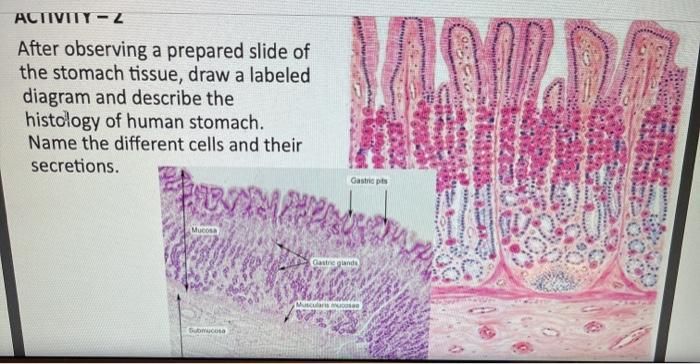
Solved ACTIVITY - Z After observing a prepared slide of the ...
30 Histology Quizzes Online, Trivia, Questions & Answers - ProProfs A) TSH is derived from the hypothalamus. B) TSH is stored in the pars nervosa of the pituitary gland. C) Herring bodies store oxytocin and FSH. D) Somatotrophs are acidophils and they produce GH. E) Chromophobes secrete oxytocin and vasopressin. Sample Question. Body temperature = 98 degrees F heart rate = 80 beats/min mean arterial blood ...

MED AID INDIA Oral Histology Prepared Glass Slide, for ...
Epididymis histology slide diagram and identification points In the epididymis histology slide labeled diagrams, you will find all the characteristics features. If you need more related diagrams of an epididymis slide, please find these here . You may also identify the following slides - The vas deference histology slide with a labeled diagram A seminal vesicle histology slide and its identification points
Histology Slides 1
Adrenal gland histology slide diagram and identification points All the histological features are shown in the adrenal gland slide labeled diagram. You will get more diagrams here. Suggested slides for you - The thyroid gland histology slide with a labeled diagram and identification points Identification of the pituitary gland slide and drawing the diagram Adrenal gland histology slide image drawing
Biology W2501 :: Contemporary Biology Lab -- Histology
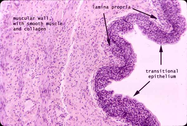
Pathology Outlines - Anatomy & histology Hilar cell hyperplasia: associated with hCG administration, pregnancy and choriocarcinoma. Rete ovarii: Counterpart of rete testis. Seen as clefts, tubules, cysts, papillae lined by cuboidal or columnar epithelium. Located usually in the hilum of the ovary, surrounded by spindle cell stroma. Walthard cell nests:

Histology Slides
Anatomy, Histology, & Pathology | Continuing Studies Investigate the appearance of cells in various organ systems, including cardiovascular, digestive, respiratory, reproductive and nervous. After seeing the normal tissue state, you will explore the appearance of cells in various disease states (pathology), such as bone osteoporosis, myocardial heart infarction, neoplasms and more.

Shotgun Histology Epiglottis
Histology - An Essential Textbook PDF Free Download Learn to identify histological structures and their correlated functions! Histology: An Essential Textbook is a concise, multimedia study guide for medical students who need to learn the functions and related correlations of cells, tissues, and organs of the human body. Professor D.J. Lowrie, Jr. has written a unique and practical medical ...
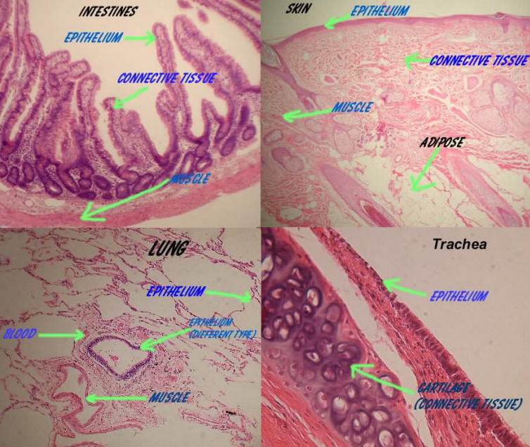
Digestive
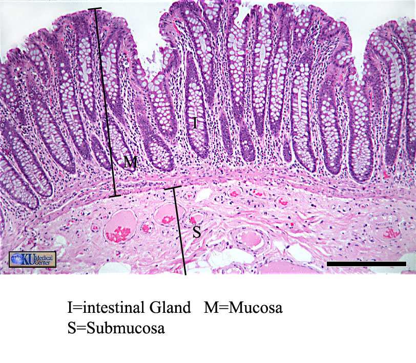
Colon Histology Slide with Labeled Diagram - AnatomyLearner Colon Histology Slide with Labeled Diagram 04/06/2022 04/06/2022 by anatomylearner The colon histology slide possesses the typical four layers of a tubular organ - mucosa, submucosa, muscularis, and serosa. But, there are no permanent plica circularis and villi in the colon slide as found in the different segments of the small intestine.

Bresser | BRESSER Prepared Slides: Histology | Expand Your ...

Unit 4 - Lab 1 & 3 Histology Slides Flashcards | Quizlet

Human anatomy and physiology, Histology slides, Medical ...
Histology at SIU
Epithelia: The Histology Guide
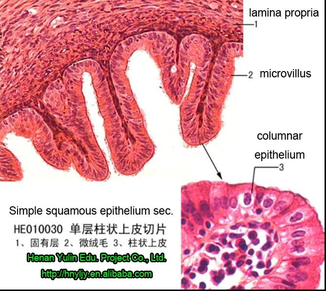
histological slides,simple columnar epithelium of Human sec ...

Using Histology Slides to Enhance Mammalian Dissection ...
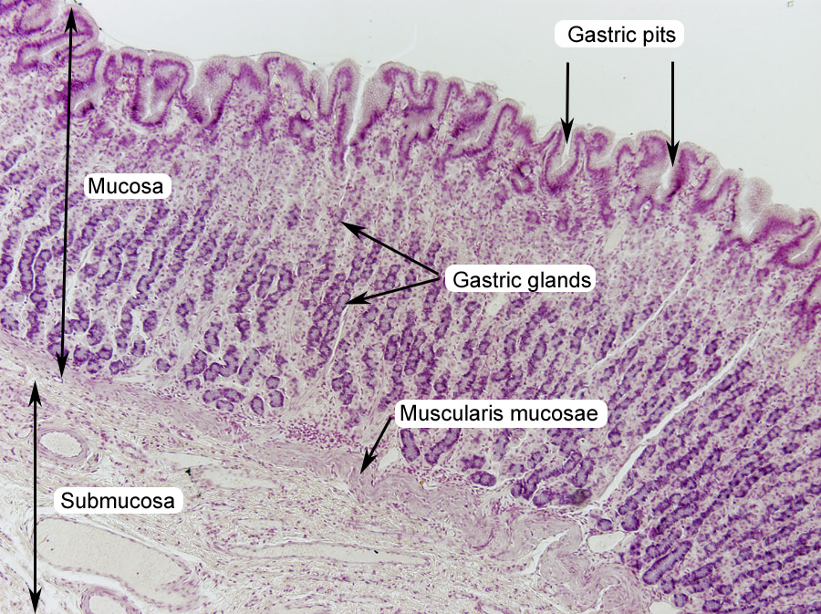
Pharynx, Esophagus, and Stomach | histology
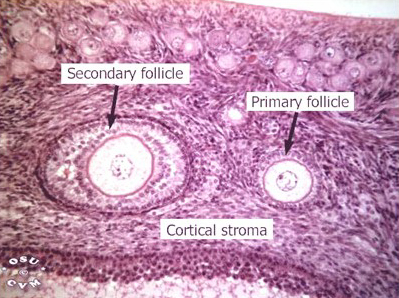
Ovary Histology - Ovary (labels) - histology slide -

583 Histology Slides Photos and Premium High Res Pictures ...
Histology Tutor
Histology Slides at Thomas Scientific

Prepared Microscope Slide - Virtual Histology - Sebaceous ...
Biology W2501 :: Contemporary Biology Lab -- Histology
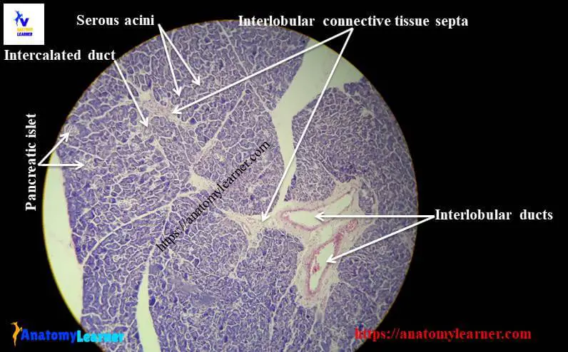
Pancreas Histology - Identifying Features with Labeled Slide ...
Liver histology: Structure, cells and characteristics | Kenhub
Histology slides snapshots (first year mbbs)
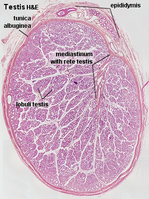
HA235 - Histology - Male Reproductive System

Quiz - Histology@Yale
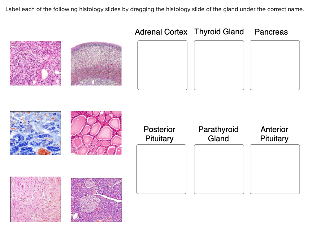
Solved Label each of the following histology slides by ...

ovary-slide-labelled-histology | SchoolWorkHelper
Histology slide guide

Micro Histology Slides, Tissue Microscope Slides -Alibaba.com

Integumentary System Histology - Skin (labels) - histology ...

Basic Histology of the Eye and Accessory Structures - EyeWiki

Histology of Blood Vessels
Post a Comment for "40 labeled histology slides"