39 labelled sarcomere
IB Questionbank 15N.2.HL.TZ0.3a(i): Label the structures indicated on the X-ray of a human elbow. 15N.2.HL.TZ0.6a: Draw a labelled diagram to show the structure of a sarcomere. 15N.3.SL.TZ0.5a: Draw a labelled diagram showing the arrangement of proteins in a sarcomere. 13M.1.HL.TZ1.37: What is the role of ATP during contraction of a skeletal muscle fibre? A. Skeletal muscle - Wikipedia WebSkeletal muscles (commonly referred to as muscles) are organs of the vertebrate muscular system and typically are attached by tendons to bones of a skeleton. The muscle cells of skeletal muscles are much longer than in the other types of muscle tissue, and are often known as muscle fibers. The muscle tissue of a skeletal muscle is striated – having a …
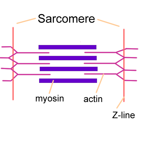
Sarcomere - an overview | ScienceDirect Topics A sarcomere is the basic contractile unit of muscle fiber. Each sarcomere is composed of two main protein filaments—actin and myosin—which are the active structures responsible for muscular contraction. The most popular model that describes muscular contraction is called the sliding filament theory.

Labelled sarcomere
Labeled Sarcomere Diagram Start studying Sarcomere Labeling. Learn vocabulary, terms, and more with flashcards, games, and other study tools. A sarcomere is the basic unit of striated muscle tissue. It is the repeating unit between two Z lines. Skeletal muscles are composed of tubular muscle cells which. Sarcomeres are composed of thick filaments and thin filaments. Sarcomere- Definition, Structure, Diagram, and Functions A sarcomere is a complex multicomponent biological system and functional unit of striated muscle which plays a vital role in transforming the chemical energy released upon the ATP hydrolysis into mechanical work. Skeletal muscles are made up of the basic unit called a sarcomere and all voluntary movement is initiated by this skeletal muscle. Sarcomere Labeling Quiz - PurposeGames.com Sarcomere Labeling by emcanallen 52,724 plays 8 questions ~ 20 sec More 16 5.00 (you: not rated) Language English Tries Unlimited [?] Last Played February 22, 2022 - 12:00 am There is a printable worksheet available for download here so you can take the quiz with pen and paper. Remaining 0 Correct 0 Wrong 0 Press play! 0% 10:00.0 Highscores
Labelled sarcomere. Myofibril - Components, Appearance, Structure, Function and … WebAlso, make sure the diagram is properly labelled. The skeletal muscle cells are long and cylindrical, and they are also referred to as the muscle fibres or myofibers. The skeletal muscle fibres are very large when compared to other cells. They have lengths up to 30 cm and a diameter which is up to 100 micrometres, and this is the example of the sartorius of … Actin - Wikipedia WebActin is a family of globular multi-functional proteins that form microfilaments in the cytoskeleton, and the thin filaments in muscle fibrils.It is found in essentially all eukaryotic cells, where it may be present at a concentration of over 100 μM; its mass is roughly 42 kDa, with a diameter of 4 to 7 nm.. An actin protein is the monomeric subunit of two types of … HBS - Labeling a Sarcomere Quiz - Quizizz Question 5. 300 seconds. Q. In a contracted muscle fiber, identify what occurs within the following area of the sarcomere - sarcomere length. answer choices. increase. decrease. stay the same. Question 6. The Sarcomere and Sliding Filaments in Muscular Contraction: Definition ... Striated Muscle Arrangement. Before we examine the contents of an individual sarcomere, let's take a look at how sarcomeres are arranged within the context of a striated muscle cell - that is, a ...
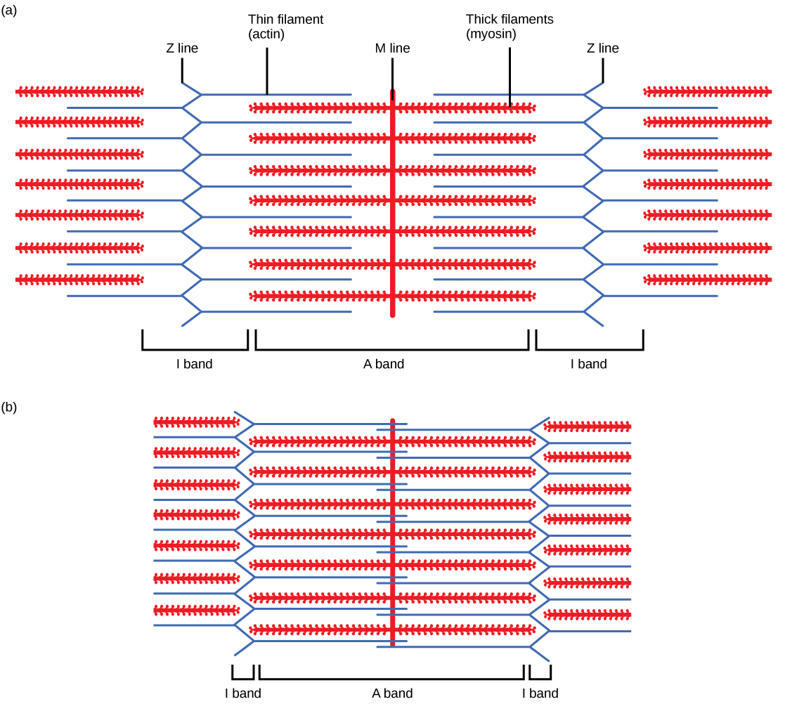
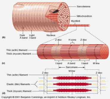
Sarcomere: Structure and Parts, Functions and Histology Sarcomere: Structure and Parts, Functions and Histology A sarcomere it is the fundamental functional unit of striated muscle, that is, of skeletal and cardiac muscle. Skeletal muscle is the type of muscle that is used in voluntary movement and the heart muscle is the muscle that is part of the heart. Which of the following sarcomeres is labelled correctly A sarcomere is composed of thick filaments made up of actin and myosin, respectively. A-band is the region where thick filaments are present. In the H-zone only thin filaments are present. I bands are the regions where thin bands are exclusively present. The Z-lines represent the boundaries between different sarcomeres. Sarcomere Photos and Premium High Res Pictures - Getty Images Browse 26 sarcomere stock photos and images available, or search for myofibril or epimysium to find more great stock photos and pictures. of 1. NCERT Solutions for Class 11 Biology Chapter 20 Locomotion ... 3. Contraction of sarcomere occurs when the actin filaments are pulled in the opposite ends. 4. During the process of contraction, the I-band shortens whereas the A-band maintains its length causing the muscles to contract. 4. Write true or false. If false change the statement so that it is true. (a) Actin is present in thin filament
Sliding Filament Theory - Definition, Diagram and Important FAQs The sliding filament theory is given by A. F. Huxley and R. Niedergerke (1954), and H. E. Huxley and J. Hanson (1954) explain how muscles in the human body contract to produce force.). In 1954, using high-resolution microscopy, these scientists noticed changes in the sarcomeres as muscle tissue shortened. They observed that during contraction ... Physiology Multiple Choice Question Bank - Academia.edu WebThe coding letters (from a to k) within the square brackets [ ] after the question code indicate which paper(s) the question was on. The key is: a = Mar 96 paper b = Jul 96 paper c = Mar 97 paper d = Jul 97 paper e = Mar 98 paper f = Jul 98 paper g = Mar 99 paper h = Jul 99 paper i = Feb 00 paper j = Jul 00 paper k = Apr 01 paper A. 1. DRAW/ PASTE labelled HISTOLOGICAL features of | Chegg.com 3. Draw/ paste labelled sarcomere structure. 4. Define the agonists/antagonists with respect to muscle actions. 5. Using the sarcomere structure as a reference, explain how the various band's (A, H, I) length changes with contraction. (Use diagram, one relaxed sarcomere and one contracted sarcomere to support your explanation.) 6. Solved Label the skeletal muscle cell structures and - Chegg Question: Label the skeletal muscle cell structures and features of the sarcomere. Z disk M line Thick filaments Sarcomere Myofibril I band A band Sarcoplasmic reticulum Thin filaments Skeletal muscle fiber This problem has been solved! See the answer Show transcribed image text Expert Answer 100% (8 ratings)
Sarcomere - an overview | ScienceDirect Topics
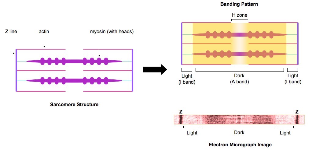
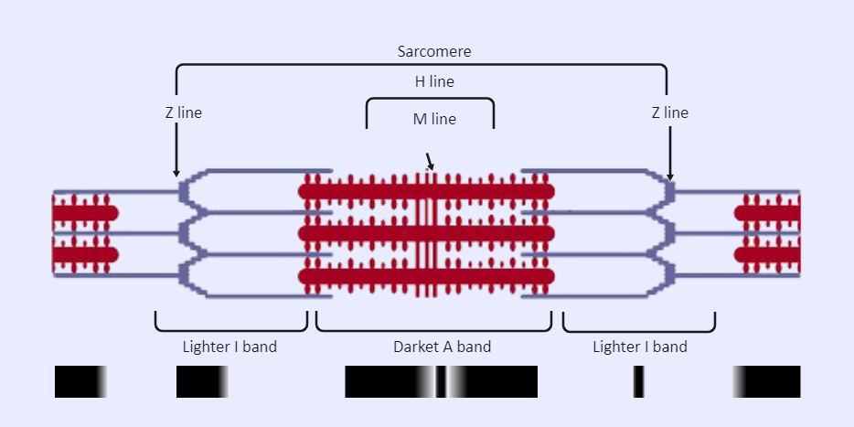
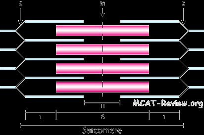
Sarcomeres: "I" and "A" Bands, "M" and "Z" Lines, "H" Zone • A sarcomere is the basic contractile unit of skeletal muscle that is made of thick and thin filaments. • Thick filaments are organized bundles of myosin, while thin filaments are made of actin along with the two other regulatory proteins-troponin and tropomyosin. • Z-lines define the boundaries of each sarcomere.

295 Sarcomere Images, Stock Photos & Vectors | Shutterstock
Sarcomere - Definition, Structure, Function and Quiz - Biology Dictionary A sarcomere is the functional unit of striated muscle. This means it is the most basic unit that makes up our skeletal muscle. Skeletal muscle is the muscle type that initiates all of our voluntary movement. Herein lies the sarcomere's main purpose. Sarcomeres are able to initiate large, sweeping movement by contracting in unison.

Draw the Diagram of a Sarcomere of Skeletal Muscle Showing ...
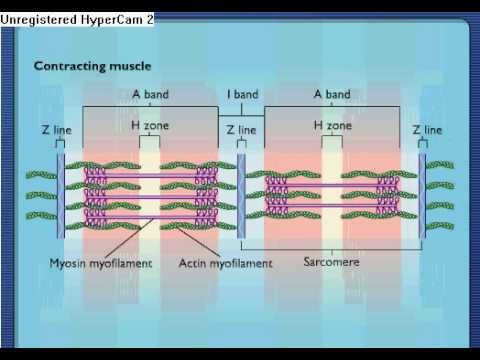
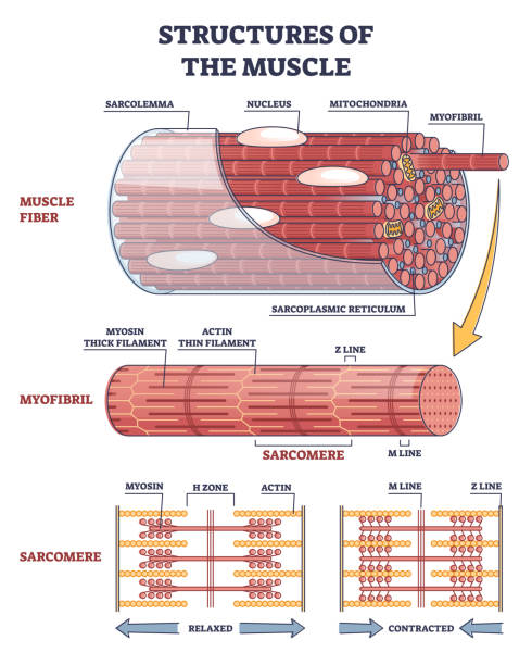
What Is the Difference Between Relaxed and Contracted Sarcomere? Groups of thick and thin filaments that alternately overlap and move apart are called sarcomeres. The areas between the thick and thin filaments during a relaxed state are called I bands, H zones and A bands. During contraction and relaxation, the length of the filaments remains the same. Myosin heads grasp the thin filaments and either push or ...
B1 Muscles and Movement | Brent Cornell
Comparison of a relaxed and contracted sarcomere (A) The basic organization of a sarcomere subregion, showing the centralized location of myosin (A band). Actin and the z discs are shown in red. (B) A conceptual diagram representing the...
What is the relationship between muscle contraction and ATP ...
Label the Sarcomere Quiz - PurposeGames.com This is an online quiz called Label the Sarcomere There is a printable worksheet available for download here so you can take the quiz with pen and paper. From the quiz author Structure of a Skeletal Muscle Sarcomere Your Skills & Rank Total Points 0 Get started! Today's Rank -- 0 Today 's Points One of us! Game Points 17

Sarcomere location of MyBP-C. The electron micrograph was ...
Sarcomere (Muscle) Coloring - The Biology Corner The enter muscle fiber is surrounded by the sarcolemma (D), color this membrane brown. If expanded, the light and dark bands are shown as individual thick and thin filaments. Color the thick filaments (not labeled) red and the thin filaments blue. The Z line is the boundary between sarcomeres, named after its shape. Color the Z-line orange.

muscle-fiber.html 50_25bSarcomereStructure-L.jpg
A general strategy to develop cell permeable and fluorogenic ... - Nature Web02.12.2019 · f, No-wash live-cell confocal images of co-cultured normal U2OS cells and U2OS FlpIn Halo-SNAP-NLS-expressing cells labelled with 500 nM 29 (left) or 33 (right) for 30 min. g, Structures of SiR700 ...

15. Muscular System: The Sarcomere and Myofilaments - LabXchange
Sarcomere | Definition, Structure, & Sliding Filament Theory The sarcomere is the basic unit function with muscle fiber cells. This is a distinguishing unit in some types of muscle tissue. Due to the striated nature of both skeletal muscle and cardiac muscle is observed by microscope slides. Myofibril: Myofibril is a very fine contractile muscle fiber cells.
19.4 Muscle Contraction and Locomotion – Concepts of Biology ...
Cells of the adult human heart | Nature Sep 24, 2020 · Cardiomyocytes show high-level expression of genes that encode contractile force-generating sarcomere proteins (TTN, MYBPC3 and TNNT2) and ... Each cellular compartment was labelled under the ...

Draw a neat labelled diagram of a sarcomereof skeletal muscle ...
Label the Sarcomere Structure Diagram | Quizlet Start studying Label the Sarcomere Structure. Learn vocabulary, terms, and more with flashcards, games, and other study tools.
Sarcomere Labeled | EdrawMax Template
Guide design resources — Zhang Lab WebThank you to the thousands of users who visited our guide design tool over the past five years. We recently shut down crispr.mit.edu, but there are many other guide design tools available that we hope you will find helpful.
MOVEMENT
Browse Articles | Nature Web15.12.2022 · Browse the archive of articles on Nature. Msemburi et al. describe how the World Health Organization has estimated the excess mortality associated with the COVID-19 pandemic, by month and for 2020 ...
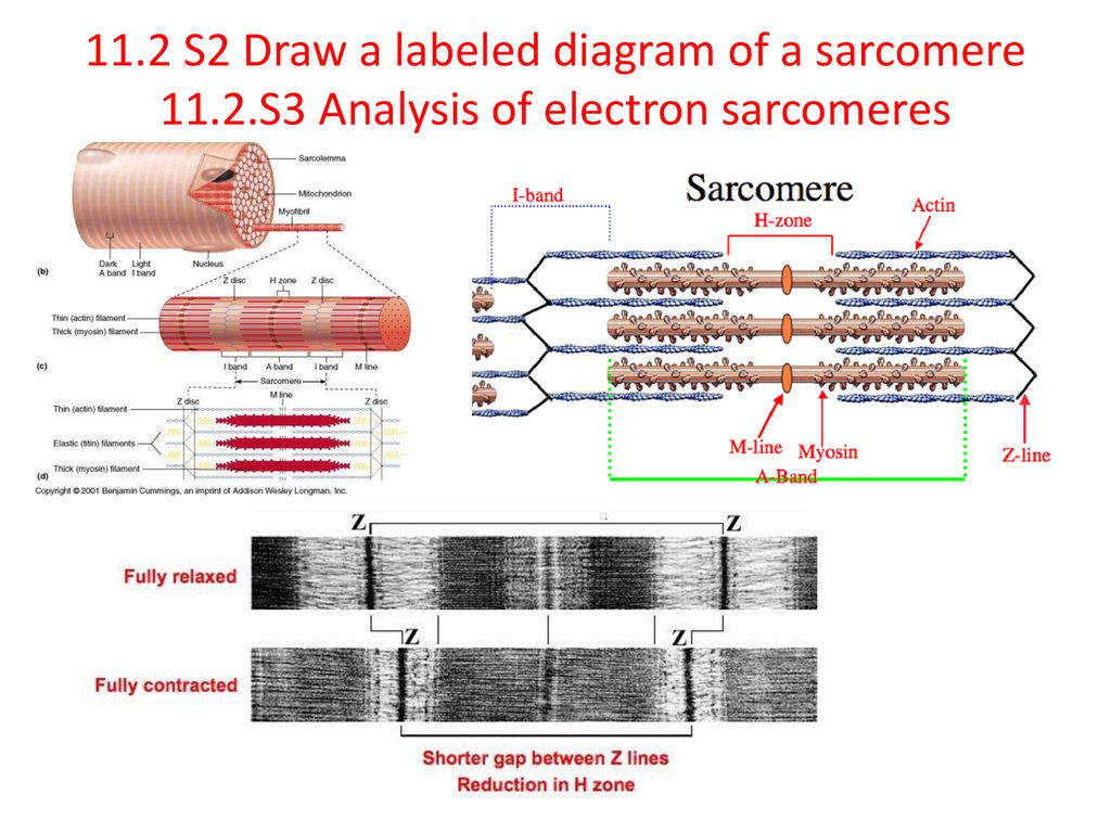
11.2 Movement. - ppt download
art-labeling activity: sarcomere structure - 4-h-dairy-posters Diagram and label a sarcomere including a thick filament thin filament A band H zone I band Z A. However thick and thin filamentsthe components of sarcomeresdo not shorten. Skin that has four layers of cells is referred to as thin skin From deep to superficial these layers are the. Solved Art Labeling Activity Sarcomere Structure Drag The Chegg ...

Name the regions of the sarcomere labelledD E and F in the ...
Physical Education MCQ Questions - examyear.com Oct 30, 2021 · 30. Given below are two statements, one labelled as Assertion (A) and the other labelled as Reason (R). Assertion (A) : Physical Education is an art. Reason (R) : Performing gymnastic activities has an aesthetic and creative value. In the context of the above two statements, which one of the following is correct? (A) (A) is right, but (R) is wrong.
Solved 1. Label the diagram of the Sarcomere below: n'-' 2 ...
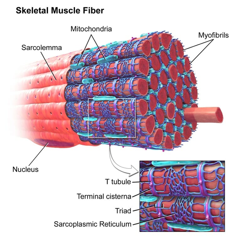
T-tubule - Wikipedia T-tubules (transverse tubules) are extensions of the cell membrane that penetrate into the center of skeletal and cardiac muscle cells.With membranes that contain large concentrations of ion channels, transporters, and pumps, T-tubules permit rapid transmission of the action potential into the cell, and also play an important role in regulating cellular calcium concentration.
Biology 2404 A&P Basics
Cardiac muscle physiology | BJA Education | Oxford Academic Web03.05.2007 · Cross bridge cycling occurs, leading to a shortening of the sarcomere and resultant muscular contraction. As intracellular calcium concentrations decrease during repolarization, calcium dissociates from troponin as intracellular calcium concentration decreases, resulting in relaxation. Diastolic relaxation is an active (ATP-dependent) …
IJMS | Free Full-Text | Current Understanding of Residual ...
UNIT 5: Label the parts of the Sarcomere Flashcards | Quizlet the line formed by the attachment of actin filaments between two sarcomeres of a muscle fiber in striated muscle cells WHERE IS THE Sarcomere? BC WHERE IS THE A Band? AC Dark band Actin and myosin filaments are both present in this dense region WHERE IS THE Myosin MYOFILAMENT? BD I Band AB region containing thin filaments only Students also viewed

Sarcomere Diagram | Quizlet
Sarcomere: anatomy, structure and function | Kenhub The sarcomere is the main contractile unit of muscle fiber in the skeletal muscle. Each sarcomere is composed of protein filaments ( myofilaments) that include mainly the thick filaments called myosin, and thin filaments called actin. The bundles of myofilaments are called myofibrils .

Illustrate sarcomere with a diagram.
What is a Sarcomere? - Parts & Contraction - Study.com The first known use of sarcomere was in the year of 1891. It was used to define the contractile unit in a striated muscle comprised of both thick and thin filaments. Bands The A-band contains...
Sarcomere Contraction - Process Of Muscle Contraction With Myosin & Actin
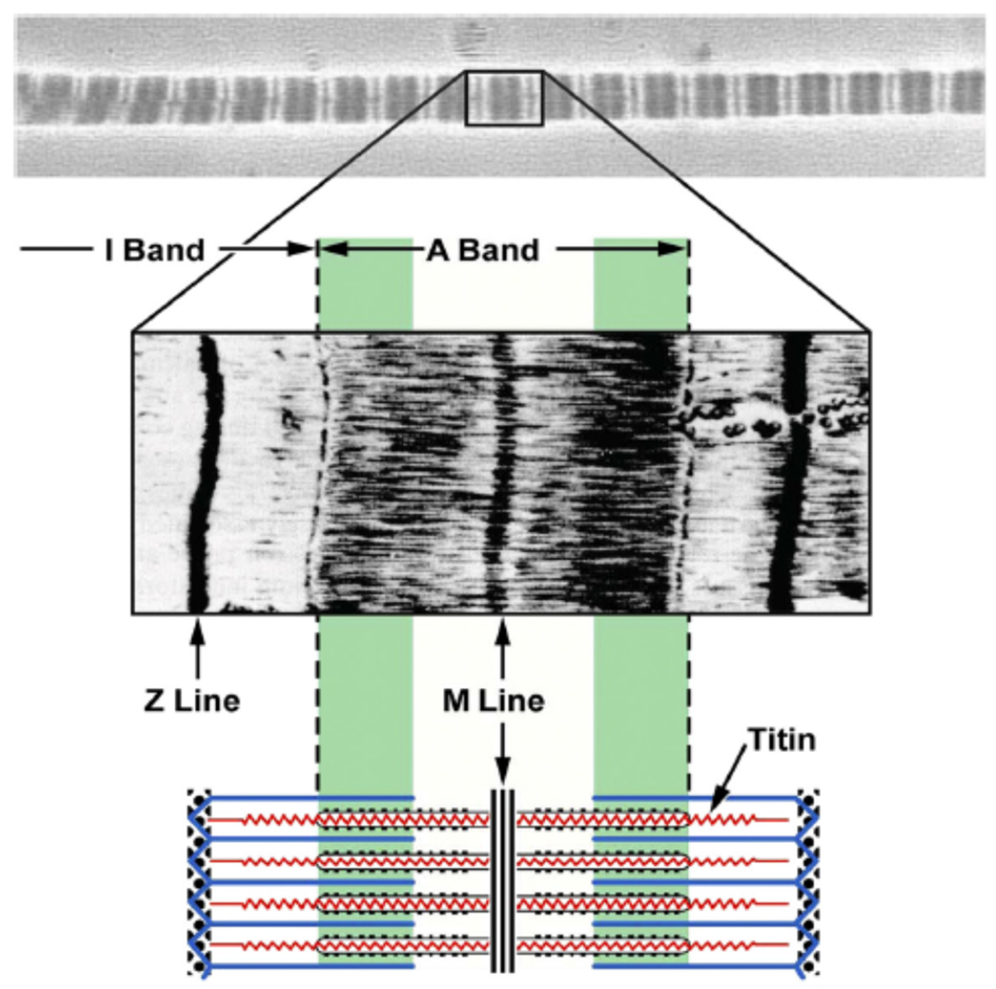
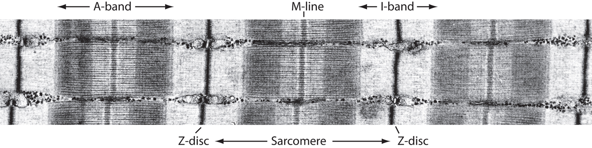
Sarcomere structure. A) Electron microscope image revealing the ... The sarcomere defines one elementary contractile unit (Fig. 7). The alternation between the A and I bands is used to estimate sarcomere length. Due to their size and force ranges, researchers have ...

Muscle tissue - Knowledge @ AMBOSS
Diagram Of Sarcomere A sarcomere is the basic unit of striated muscle tissue. It is the repeating unit between two Z lines. Skeletal muscles are composed of tubular muscle cells which. sarcomere. Schematic: The Z line is depicted in black, myosin in red, actin in green/gray, and tropomyosin in blue. Image: MPI of Molecular Plant Physiology. Sarcomere definition.
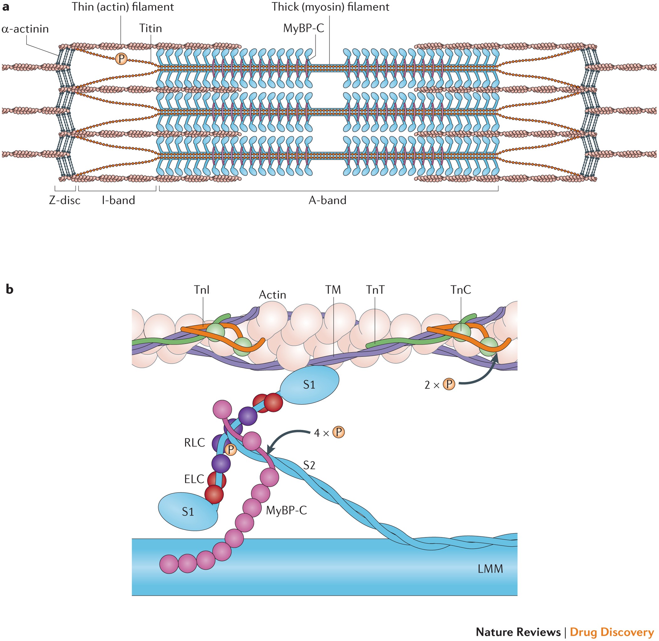
Targeting the sarcomere to correct muscle function | Nature ...
10.2 Skeletal Muscle - Anatomy & Physiology The sarcomere is the smallest functional unit of a skeletal muscle fiber and is a highly organized arrangement of contractile, regulatory, and structural proteins. It is the shortening of these individual sarcomeres that lead to the contraction of individual skeletal muscle fibers (and ultimately the whole muscle).
Structures Of Muscle With Fiber Myofibril And Sarcomere ...
Sarcomere - Wikipedia A sarcomere (Greek σάρξ sarx "flesh", μέρος meros "part") is the smallest functional unit of striated muscle tissue. [1] It is the repeating unit between two Z-lines. Skeletal muscles are composed of tubular muscle cells (called muscle fibers or myofibers) which are formed during embryonic myogenesis. Muscle fibers contain numerous ...

Given below is the figure of a sarcomere. Identify the parts ...
Sarcomere Labeling Quiz - PurposeGames.com Sarcomere Labeling by emcanallen 52,724 plays 8 questions ~ 20 sec More 16 5.00 (you: not rated) Language English Tries Unlimited [?] Last Played February 22, 2022 - 12:00 am There is a printable worksheet available for download here so you can take the quiz with pen and paper. Remaining 0 Correct 0 Wrong 0 Press play! 0% 10:00.0 Highscores

The Sarcomere and Sliding Filaments in Muscular Contraction ...
Sarcomere- Definition, Structure, Diagram, and Functions A sarcomere is a complex multicomponent biological system and functional unit of striated muscle which plays a vital role in transforming the chemical energy released upon the ATP hydrolysis into mechanical work. Skeletal muscles are made up of the basic unit called a sarcomere and all voluntary movement is initiated by this skeletal muscle.

0779-00 Complete Sarcomere Model – Denoyer-Geppert Science ...
Labeled Sarcomere Diagram Start studying Sarcomere Labeling. Learn vocabulary, terms, and more with flashcards, games, and other study tools. A sarcomere is the basic unit of striated muscle tissue. It is the repeating unit between two Z lines. Skeletal muscles are composed of tubular muscle cells which. Sarcomeres are composed of thick filaments and thin filaments.

Sarcomere Structure Tutorial | Sophia Learning | Human muscle ...

Sarcomeres I And A Bands M And Z Lines H Zone - Specialized ...

Sarcomere Labeling Diagram | Quizlet

Muscles

38.16: Muscle Contraction and Locomotion - Sliding Filament ...

Which of the following sarcomeres is labelled correctly ...
2. Pada otot lurik ada dua protein penyusunnya yai...

Given below is the figure of a sarcomere. Identify the parts ...

Identifying Regions in the Sarcomere
Sarcomere - Definition, Structure, Function and Quiz ...
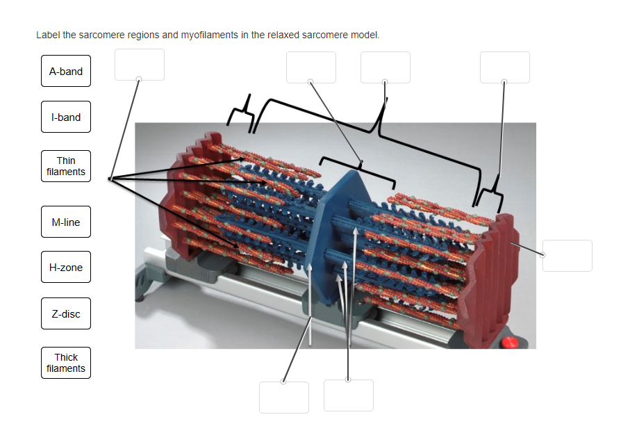
Solved Label the sarcomere regions and myofilaments in the ...
Structure and Composition of Muscle - Meat Science
The Titin/Telethonin Complex
Post a Comment for "39 labelled sarcomere"