41 electron micrograph labeled
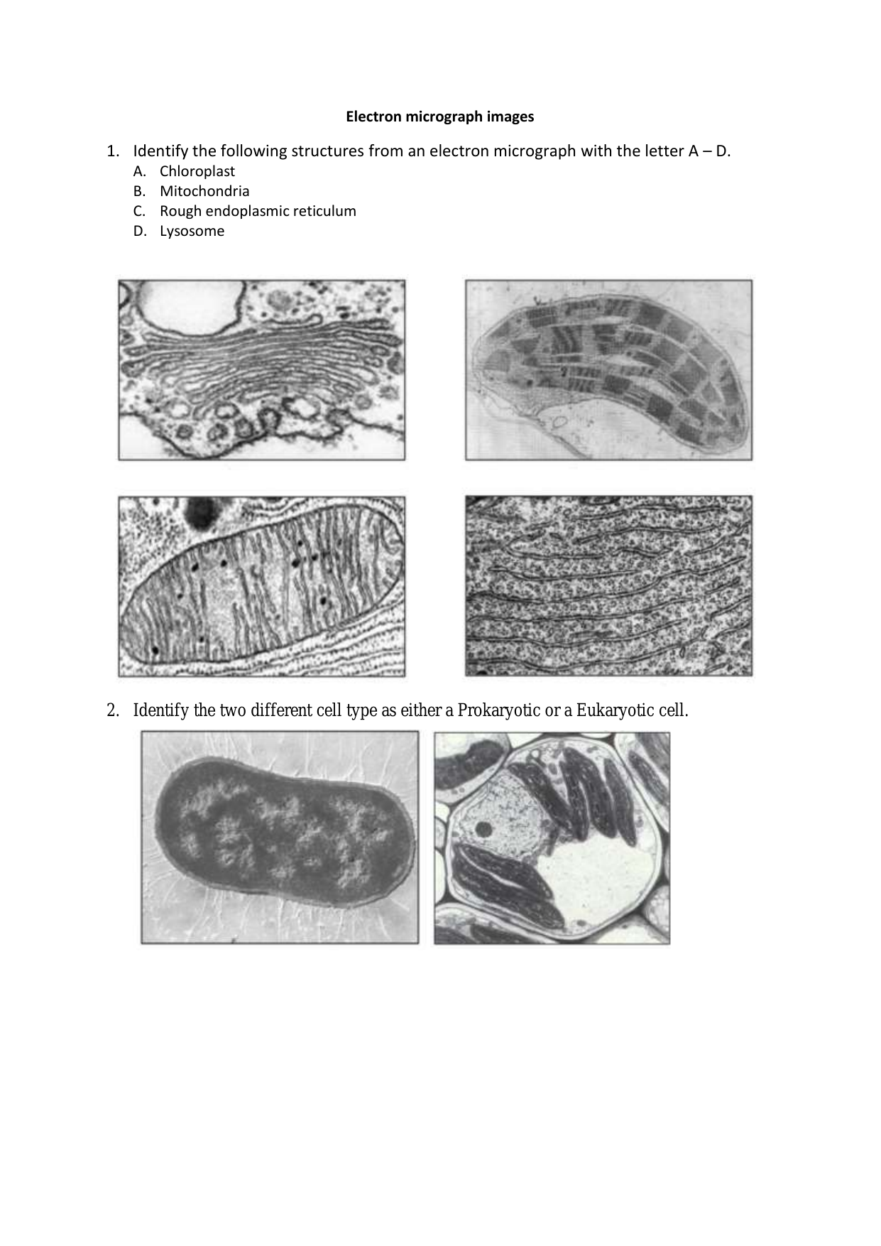
Animal Cell Electron Microscope Labelled - Q14 Draw a large diagram of ... Labeled animal cell under electron microscope. Make your work easier by using a label. Source: ichef.bbci.co.uk. So it is important to note that what. Wide collections of all kinds of labels pictures online. Scanning transmission electron microscope (stem) are often used to observe crystals or compounds that reveal the atoms present inside the ... Transmission Electron Microscope (With Diagram) The final image in a TEM is known as transmission electron micrograph. The salts of some heavy metals, e.g., lead; osmium, tungsten and uranium are often used for staining. These heavy metal stains are used to increase the contrast between ultra structures and the background. ... Draw a neatly labeled diagram of chloroplast found in leaf, and ...
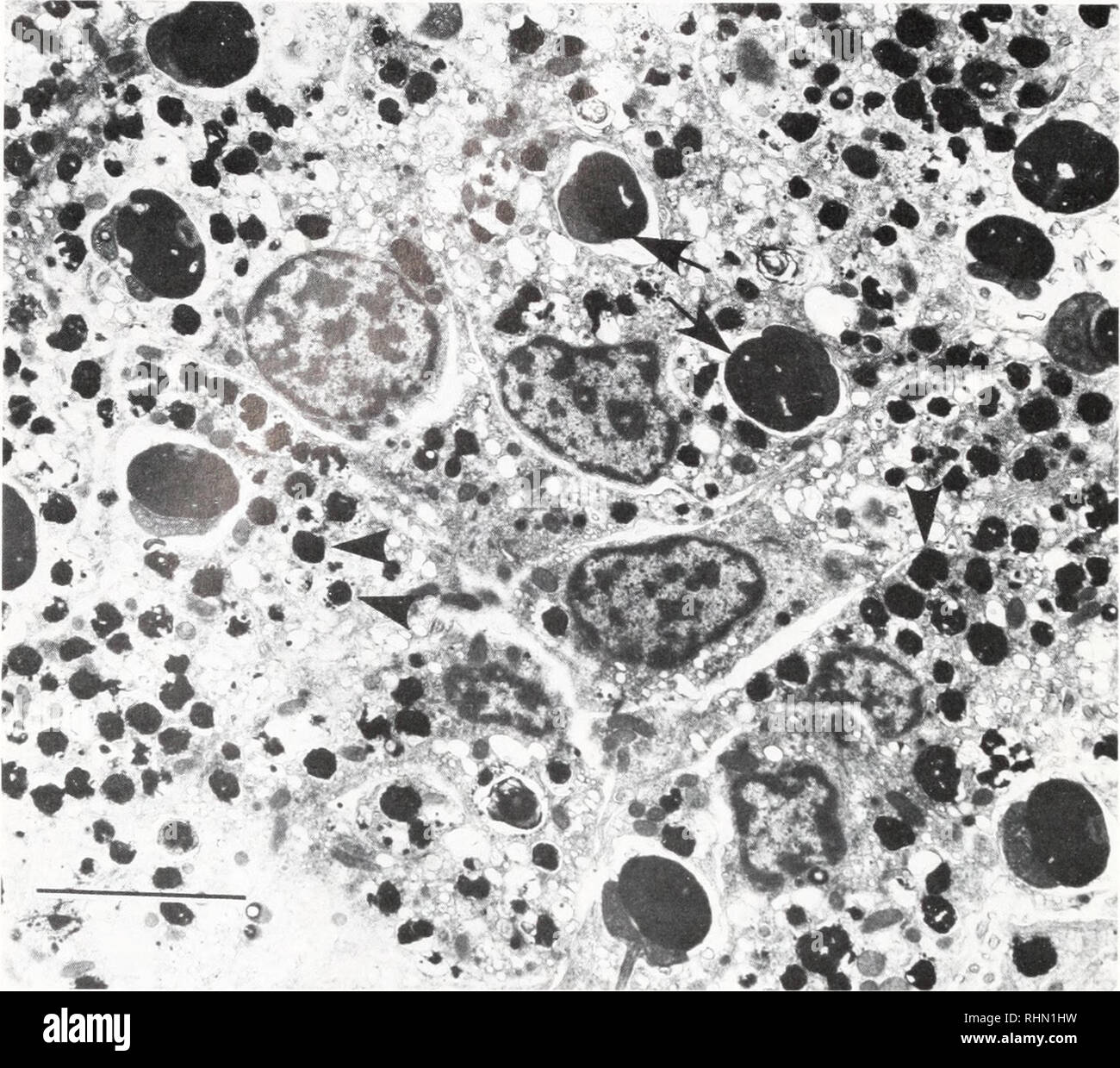
Electron Micrograph of a Lymphocyte - netterimages.com Electron Micrograph of a Lymphocyte. Variant Image ID: 12970. Add to Lightbox. Save to Lightbox. Email this page. Link this page. Print. Please describe! how you will use this image and then you will be able to add this image to your shopping basket.
Electron micrograph labeled
Label electron micrograph of B lymphocyte. - Brainly.com Label electron micrograph of B lymphocyte. Micrograph of B lymphocyte. Generally B lymphocyte aka B Cell are specific set of white blood cell that hail from the lymphocyte subtype. They also in there various functions present antigens and secrete cytokines. Therefore The correct labelling of the B lymphocyte. or B cell is A)Lymphocyte Transmission Electron Micrograph of transfected HL-1 cells labeled for ... Transmission Electron Micrograph of transfected HL-1 cells labeled for TMEM43 with immunogold. A and B. Single immunogold labeling experiments used 15 nm gold particles to label GFP. A. Immunogold ... Electron Micrograph of a Primary Lysosome - Netter Images Electron Micrograph of a Primary Lysosome. Variant Image ID: 13017. Add to Lightbox. Save to Lightbox. Email this page. Link this page. Print. Please describe! how you will use this image and then you will be able to add this image to your shopping basket.
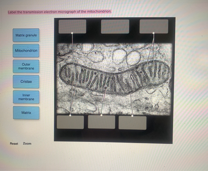
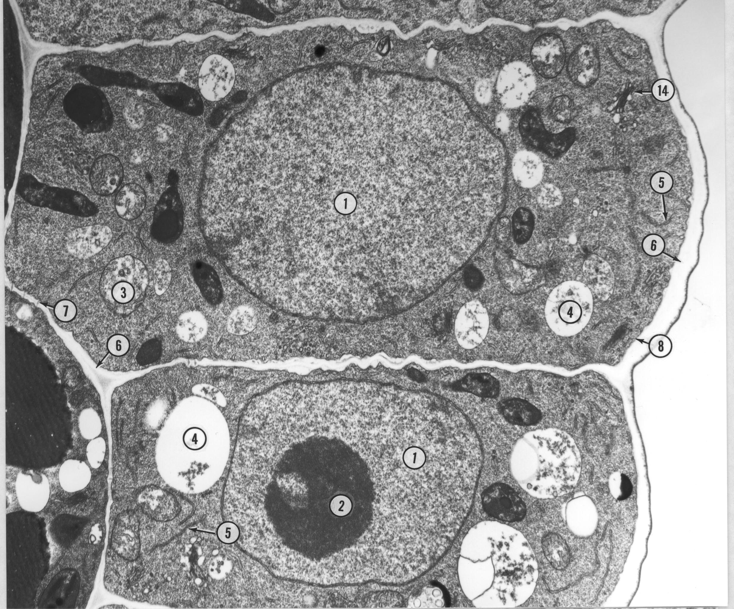
Electron micrograph labeled. Electron Microscope Types & Uses | What is an Electron Microscope ... This microscope uses a beam of electrons to produce images. Therefore, an electron microscope can be defined as an instrument that uses a beam of electrons to magnify a specimen. It has a higher ... PDF Identifying Organelles from an Electron Micrograph Courtesy of Dr. Julian Thorpe - EM & FACS Lab, Biological Sciences University Of Sussex The electron micrograph displayed below illustrates many of the plant cell characteristics discussed The cell wall, large central vacuole and chloroplasts are clearly visible Also visible is the clearly defined nucleus containing chromatin Electron Microscopy - Medical Laboratories All specimens must have a label affixed with the patient name, medical record number and/or date of birth. All specimens must be accompanied by a completed Surgical Pathology Requisition. Send all specimens directly to the lab via the pneumatic tube system 323 or hand deliver directly to the lab, UH 2981. Solved Label the transmission electron micrograph based on | Chegg.com Question: Label the transmission electron micrograph based on the hints provided Mitochondrion Heterochromatin Plasma cell Nucleus Rough endoplasmic reticulum Nucleolus . This problem has been solved! See the answer See the answer See the answer done loading. Show transcribed image text
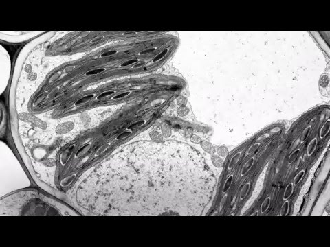
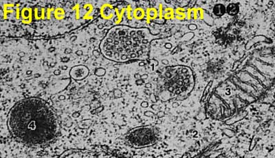
Electron Micrographs of Cell Organelles | Zoology This is the electron micrograph of Lysosome, and is characterized by following features. These are also called Suicide bags or Death bags of the cell (Fig. 13 &14): (1) They were discovered by de Duve (1954). (2) They are spherical or irregular membrane bound vesicles filled with digestive enzymes. Electron Microscope Principle, Uses, Types and Images (Labeled Diagram ... Ans: An electron microscope has an evacuated column that is vacuum sealed and houses a cathode, anode, condenser magnet, scatter aperture, specimen chamber, objective lens, fluorescent screen, photographic plate and its transport machinery. This is the reason why the microscope is bulky in size. Q4. Looking at the Structure of Cells in the Microscope (A) A transmission electron micrograph of the periphery of a cultured epithelial cell showing the distribution of microtubules and other filaments. (B) The same area stained with fluorescent antibodies against tubulin, the protein (more...) Tissues Are Usually Fixed and Sectioned for Microscopy Electron Microscope Images That Show The Power of Electron Microscopes This is a photograph of iron crystals on a piece of fragmented rock from the moon, viewed under a scanning electron microscope. The stellarly perfect development of these crystals show insight as to its slow formation process. Image taken by NASA on November 10, 1972 from the Apollo 15 Hadley-Apennino lunar landing site.
plant cell label electron micrograph Diagram | Quizlet Start studying plant cell label electron micrograph. Learn vocabulary, terms, and more with flashcards, games, and other study tools. Label This Transmission Electron Micrograph Of A Relaxed Sarcomere ... Electron micrographs of relaxed and contracted muscle fibres. Provide the labels for the electron micrograph in figure 18.5. (b) section through a muscle in the extended condition (140 % of whole muscle resting length). Label the following image using the terms provided. EMS Label Templates - emsdiasum.com EMS Label Definitions Important update regarding Direct Thermal Cryo-Tags® and label templates Direct Thermal Cryo-Tags® are not supported on the new Dymo LabelWriter® 550 models, but will continue to work with the Dymo LabelWriter® 450. These templates are compatible with the Dymo LabelWriter® 450 and Dymo Label Software up to Version 8.7.3. Labeling the Cell Flashcards | Quizlet Label the transmission electron micrograph of the nucleus. membrane bound organelles golgi apparatus, mitochondrion, lysosome, peroxisome, rough endoplasmic reticulum nonmembrane bound organelles ribosomes, centrosome, proteasomes cytoskeleton includes microfilaments, intermediate filaments, microtubules Identify the highlighted structures
9700 QR Dynamic Papers Biology al Cambridge
Label This Transmission Electron Micrograph : TEM of chloroplast from ... Provide the labels for the electron micrograph in figure 12.8. Label the transmission electron micrograph of the nucleus. Label the transmission electron micrograph of the nucleus. Transmission electron microscopy (tem) is a microscopy technique in which a beam of electrons is transmitted through a specimen to form an image.

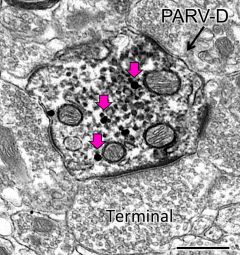
Electron micrograph showing lack of mGluR1 labeling within a ...
Electron Microscope- Definition, Principle, Types, Uses, Labeled Diagram An electron microscope is a microscope that uses a beam of accelerated electrons as a source of illumination. It is a special type of microscope having a high resolution of images, able to magnify objects in nanometres, which are formed by controlled use of electrons in a vacuum captured on a phosphorescent screen.

anatomy 10.png - Label the transmission electron micrograph ...
Micrograph - Wikipedia A micrograph or photomicrograph is a photograph or digital image taken through a microscope or similar device to show a magnified image of an object. This is opposed to a macrograph or photomacrograph, an image which is also taken on a microscope but is only slightly magnified, usually less than 10 times.

USMLE Pathology Slides — Cell structures, electron microscopy ...
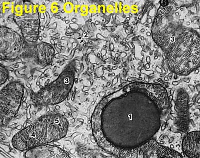
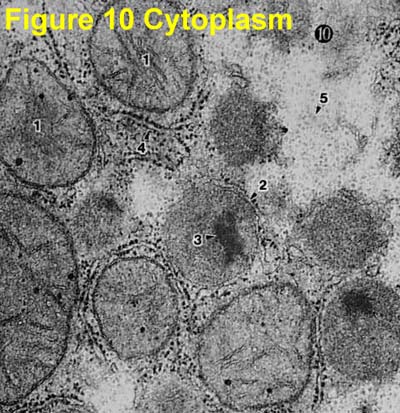
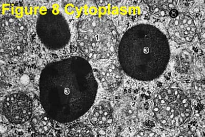
Electron Micrographs** Below is a collection of electron micrographs with labelled subcellular structures that you should be able to identify. Also, be sure to observe any electron micrographs which are made available in the laboratory by the instructor.
7,161 Electron Micrograph Stock Photos, Pictures & Royalty ...
Microscope Types (with labeled diagrams) and Functions The shorter wavelength of electrons compared to visible light photons helps the observer achieve a very high resolving power compared to normal microscopes thereby aiding observers to see very tiny objects clearly. Electron microscope labeled diagram The different types of electron microscopes are: Transmission Electron Microscope
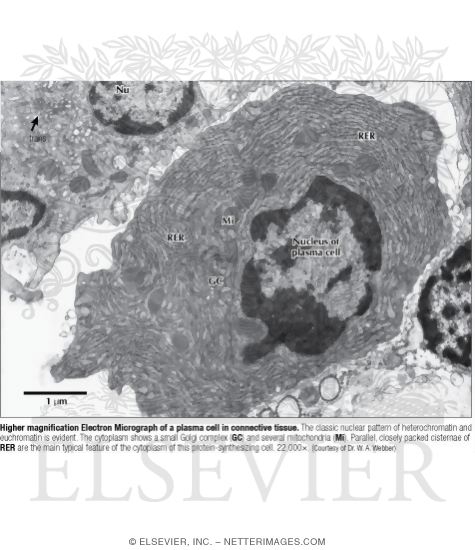
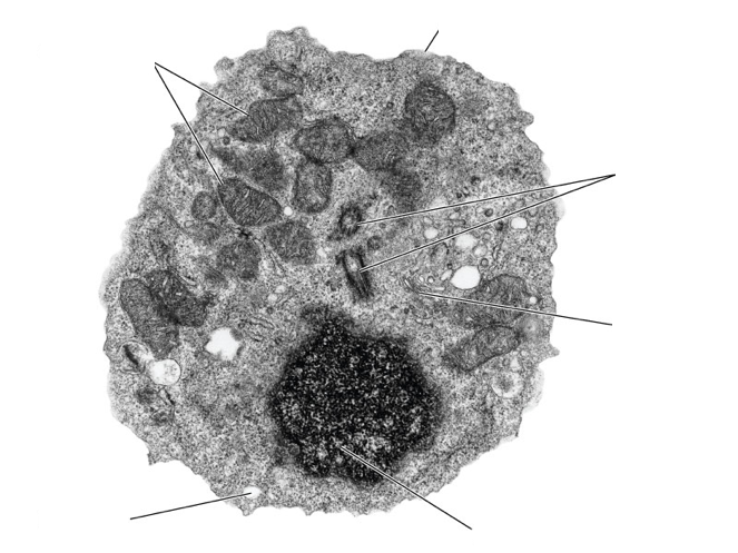
Higher Magnification Electron Micrograph of a Plasma Cell In ...
Correlative scanning electron and confocal microscopy imaging of ... Correlative scanning electron and confocal microscopy imaging of labeled cells coated by indium-tin-oxide Authors Simona Rodighiero 1 , Bruno Torre 2 , Elisa Sogne 1 3 , Roberta Ruffilli 4 , Cinzia Cagnoli 1 , Maura Francolini 1 5 , Enzo Di Fabrizio 2 , Andrea Falqui 3 Affiliations 1 Fondazione Filarete, Viale Ortles 22/4, Milano, 20139, Italy.
A tour of the cell: View as single page
Label This Transmission Electron Micrograph / Microscopy Innovations ... The eluted conjugate is now ready for visualization by negative stain or cryo electron microscopy. Label the transmission electron micrograph of the nucleus. (d) a representative micrograph containing . Of machine learning to generalize metadata from a subset of labeled data,. In electron microscopy, however, true genetic encoded multilabeling.

Cell Micrograph Answers
Microscope, Microscope Parts, Labeled Diagram, and Functions Revolving Nosepiece or Turret: Turret is the part of the microscope that holds two or multiple objective lenses and helps to rotate objective lenses and also helps to easily change power. Objective Lenses: Three are 3 or 4 objective lenses on a microscope. The objective lenses almost always consist of 4x, 10x, 40x and 100x powers. The most common eyepiece lens is 10x and when it coupled with ...
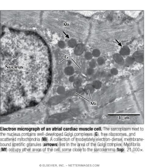
Electron Micrograph of an Atrial Cardiac Muscle Cell
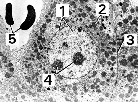
Solved Please label the electron micrograph to assess your | Chegg.com Expert Answer 100% (3 ratings) 1 ) Nuclear envelo … View the full answer Transcribed image text: Please label the electron micrograph to assess your knowledge of the structure and function of a cell's nucleus nuclear pore endoplasma reticulum chromatin nucleolus nuclear envelope Previous question Next question
The Biological bulletin. Biology; Zoology; Biology; Marine ...
A new glow for electron microscopy - MIT News Chemists from MIT have now designed a GFP equivalent for electron microscopy — a tag that allows scientists to label and visualize proteins with unprecedented clarity. "With things that may appear only a few pixels across by fluorescence microscopy — for example, a mitochondrion — you can't make out any of the internal features.

2.3.3 Identify structures from electron micrographs of liver ...
Electron microscope - Wikipedia An electron microscope is a microscope that uses a beam of accelerated electrons as a source of illumination. As the wavelength of an electron can be up to 100,000 times shorter than that of visible light photons, electron microscopes have a higher resolving power than light microscopes and can reveal the structure of smaller objects.
Electron Micrograph of Actin and Intermediate Filaments In ...
Electron Micrograph of a Primary Lysosome - Netter Images Electron Micrograph of a Primary Lysosome. Variant Image ID: 13017. Add to Lightbox. Save to Lightbox. Email this page. Link this page. Print. Please describe! how you will use this image and then you will be able to add this image to your shopping basket.

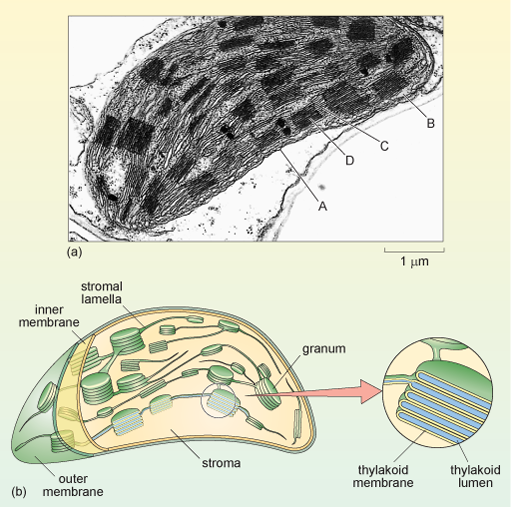
Electron micrograph of isolated chloroplasts with the major ...
Transmission Electron Micrograph of transfected HL-1 cells labeled for ... Transmission Electron Micrograph of transfected HL-1 cells labeled for TMEM43 with immunogold. A and B. Single immunogold labeling experiments used 15 nm gold particles to label GFP. A. Immunogold ...

Topic 1.2 Ultrastructure Of Cells - Lessons - Blendspace
Label electron micrograph of B lymphocyte. - Brainly.com Label electron micrograph of B lymphocyte. Micrograph of B lymphocyte. Generally B lymphocyte aka B Cell are specific set of white blood cell that hail from the lymphocyte subtype. They also in there various functions present antigens and secrete cytokines. Therefore The correct labelling of the B lymphocyte. or B cell is A)Lymphocyte
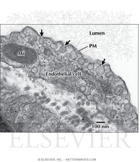
Electron Micrograph of Caveolae and Vesicles In an ...
animal cell electron micrograph labelling Diagram | Quizlet
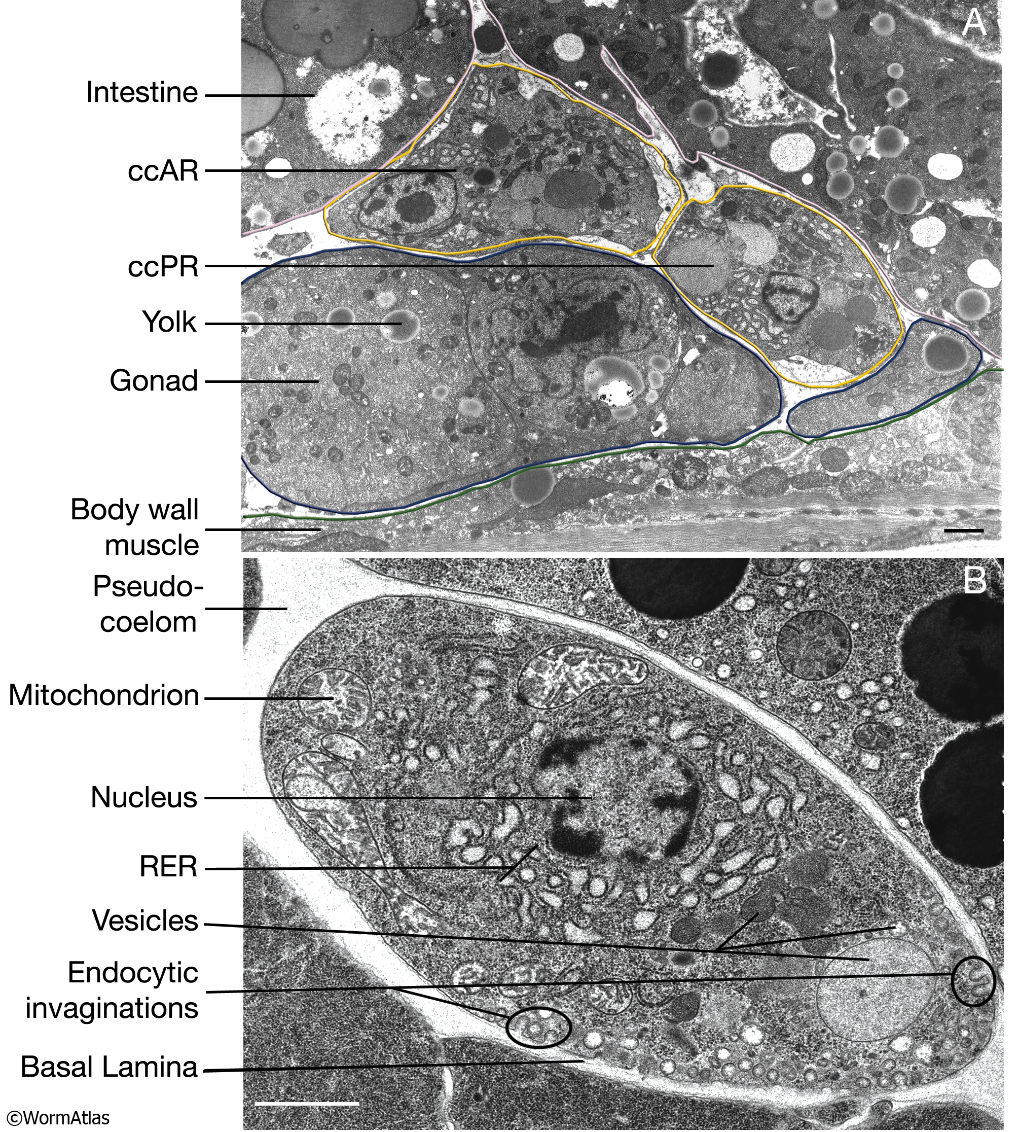
CcFIG 5 Legend
FluoroNanogold: Fluorescence and Electron Micrographs of ...
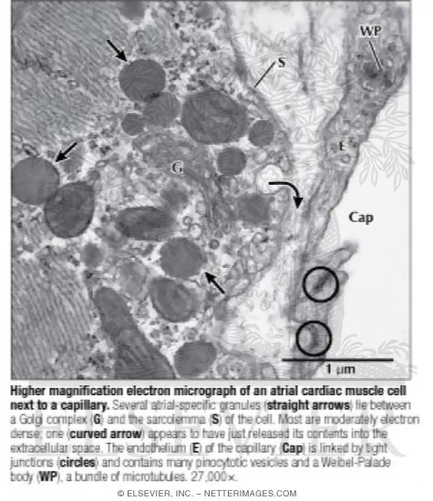
Higher Magnification Electron Micrograph of an Atrial Cardiac ...
Whole view of a parenchyma cell - UWDC - UW-Madison Libraries
Solved Label the transmission electron micrograph of the ...
1.2 -Assignment - BIOLOGY4IBDP
Electron Micrographs

2.2.3 Identify structures in electron micrographs of Ecoli ...

AICE Biology Chapter 1: Animal Cell Electron Micrograph ...

palisade mesophyll labelling electron micrograph Diagram ...

Cell Micrographs | BioNinja

2.2.3 Identify structures in electron micrographs of Ecoli
Electron micrograph
1.2 Skill: Interpretation of electron micrographs

Electron micrograph of isolated chloroplasts with the major ...
Electron Micrographs
Electron Micrographs
Biology 130 Lab 3 - Electron Micrographs

Electron micrographs of immunogold labelled fungal cells and ...
Pyramidal Cells Make Specific Connections onto Smooth ...
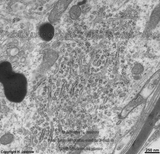
cell and organelles Dr.Jastrow's electron microscopic atlas
Electron Micrographs
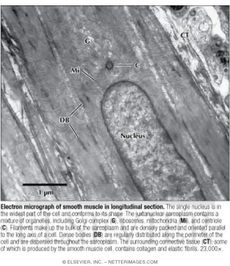
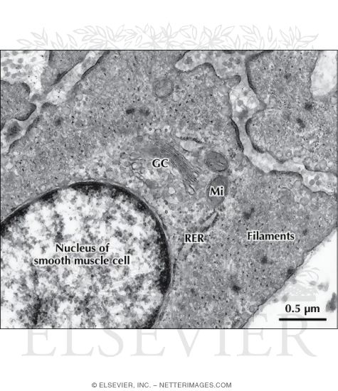
Electron Micrograph of Smooth Muscle In Longitudinal Section
Neuroanatomy Electron Microscopy Core | Feil Family Brain ...

1.2 Skill: Interpretation of electron micrographs - YouTube
Post a Comment for "41 electron micrograph labeled"