44 compound microscope drawing with label
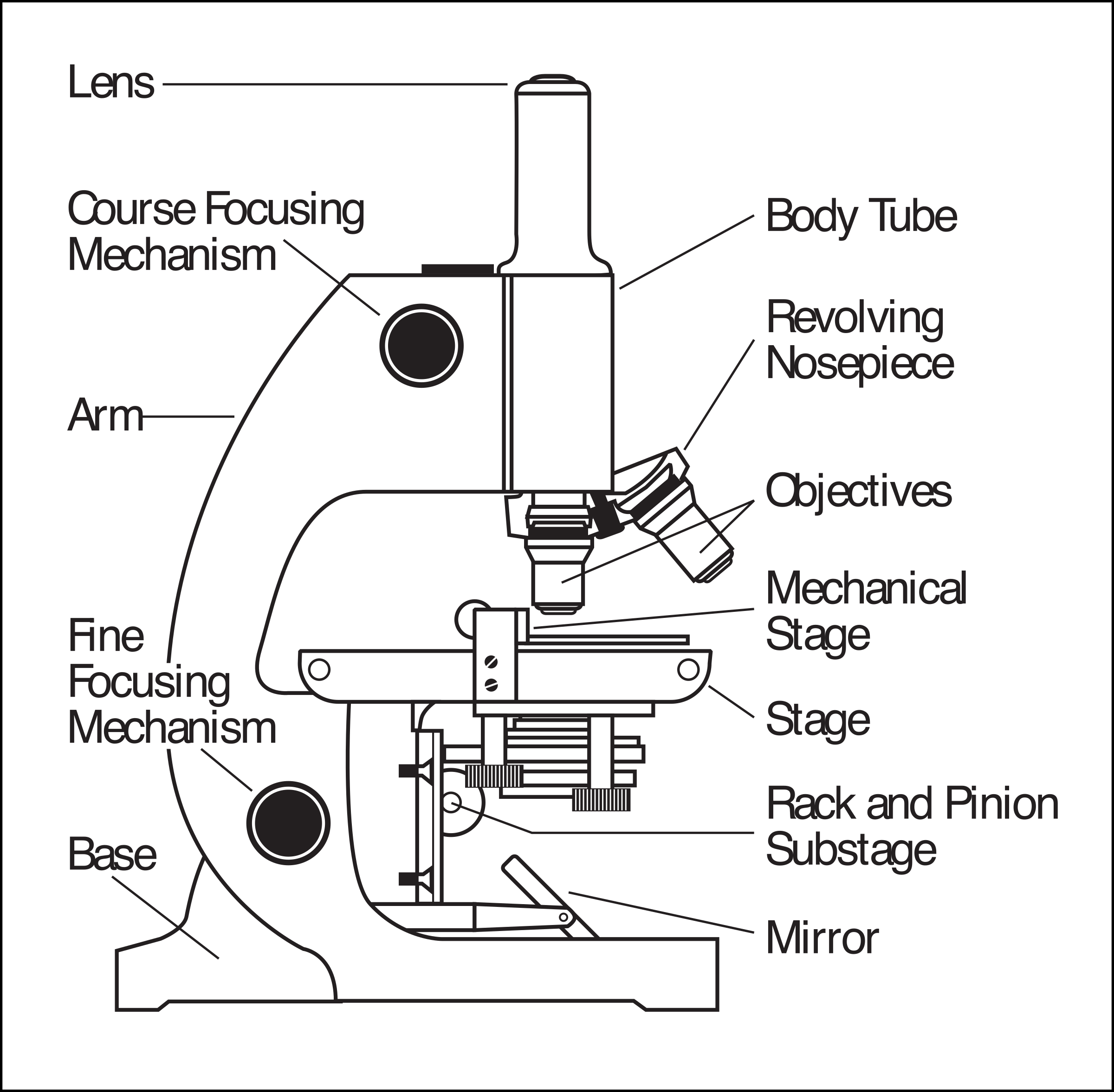
ACTIVITY-1-THE-COMPOUND-MICROSCOPE.pdf - Course Hero ACTIVITY-1-THE-COMPOUND-MICROSCOPE.pdf - Name : Rating : Course : Date : ACTIVITY 1 The Compound Microscope General Instruction: Draw and label a. ACTIVITY-1-THE-COMPOUND-MICROSCOPE.pdf - Name : Rating :... School Cotabato City State Polytechnic College; Course Title Science 123; Uploaded By MateField6213. Parts of a microscope with functions and labeled diagram Head - This is also known as the body. It carries the optical parts in the upper part of the microscope. Base - It acts as microscopes support. It also carries microscopic illuminators. Arms - This is the part connecting the base and to the head and the eyepiece tube to the base of the microscope.
Microscopy - Wikipedia The field of microscopy (optical microscopy) dates back to at least the 17th-century.Earlier microscopes, single lens magnifying glasses with limited magnification, date at least as far back as the wide spread use of lenses in eyeglasses in the 13th century but more advanced compound microscopes first appeared in Europe around 1620 The earliest practitioners of …
Compound microscope drawing with label
Draw a neat labelled diagram of a compound microscope and explain its ... Using sign convention, we find that O'I 1 = + v 0 and O'O = -u where v 0 is the image distance due to the objective and u is the object distance for the objective or the compound microscope. I 1 G 1 is negative and OJ is positive. To find me : The eyepiece behaves like a simple microscope. So : the magnifying power of the eye piece. ∴ m e ... Draw and label a compound microscope? - Answers Draw and label a compound microscope? Wiki User. ∙ 2012-09-19 09:42:29. Study now. See answer (1) Best Answer. Copy. uhmm,.... unang ana is the. 1.) Eyepiece. 2.) arm. eewan ko nakalimutan ko na ... Ark Elvin Academy Year 9 Science Study Pack Autumn … • A light microscope shines a beam of light across a thin, dead, stained specimen. • The resolution (ability to distinguish between two points) and magnification of a light microscope is high enough the view the nucleus and cell membrane. • Most organelles are too small to be viewed with a light microscope. 5. Diffusion Key information:
Compound microscope drawing with label. Compound Microscope- Definition, Labeled Diagram, Principle, Parts, Uses A compound microscope is of great use in pathology labs so as to identify diseases. Various crime cases are detected and solved by drawing out human cells and examining them under the microscope in forensic laboratories. The presence or absence of minerals and the presence of metals can be identified using compound microscopes. Isotope Distribution Calculator and Mass Spec Plotter Molecular formula of the compound of interest according to the above parameters; The title and subtitle that will appear on the graphical output; The mass scale desired in the graphic output. Both low and high mass ranges can be selected. default is 0 and 600. Compound Microscope Parts, Functions, and Labeled ... Compound Microscope Parts, Functions, and Labeled Diagram Parts of a Compound Microscope Each part of the compound microscope serves its own unique function, with each being important to the function of the scope as a whole. PharmaCircle This website uses cookies to help provide you with the best possible online experience. Please read our Terms & Conditions and Privacy Policy for information about ...
Compound Light Microscope Labeling - Printable - PurposeGames.com About this Worksheet. This is a free printable worksheet in PDF format and holds a printable version of the quiz Compound Light Microscope Labeling.By printing out this quiz and taking it with pen and paper creates for a good variation to only playing it online. 4.1. Chirality | Organic Chemistry 1: An open textbook We turn now to concept of chirality, discovered and explored by Louis Pasteur . The term chiral, from the Greek work for ‘hand’, refers to anything which cannot be superimposed on its own mirror image. Your hands, of course, are chiral – you cannot superimpose your left hand on your right, and you cannot fit your left hand into a right-handed glove (which is also a chiral object). Compound Microscope Parts – Labeled Diagram and their ... The term "compound" refers to the microscope having more than one lens. Basically, compound microscopes generate magnified images through an aligned pair of the objective lens and the ocular lens. In contrast, "simple microscopes" have only one convex lens and function more like glass magnifiers. Diagram of a Compound Microscope - Biology Discussion A bright-field or compound microscope is primarily used to enlarge or magnify the image of the object that is being viewed, which can not otherwise be seen by the naked eye. Magnification may be defined as the degree of enlargement of the image of an object provided by the microscope.
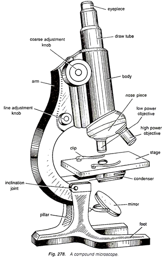
16 Parts of a Compound Microscope: Diagrams and Video In compound microscopes with two eye pieces there are prisms contained in the body that will also split the beam of light to enable you to view the image through both eye pieces. 2. Arm. The arm of the microscope is another structural piece. The arm connects the base of the microscope to the head/body of the microscope. Microscope With Labels Clip Art at Clker.com PEOPLE GOT HERE BY SEARCHING: diagrams of the microscope · light microscope and label · the compound microscope drawing · diagram of microscope with labelling ... UD Virtual Compound Microscope - University of Delaware ©University of Delaware. This work is licensed under a Creative Commons Attribution-NonCommercial-NoDerivs 2.5 License.Creative Commons Attribution-NonCommercial-NoDerivs 2.5 … Microscope Drawing And Label - Painting Valley compound parts light labeling functions microscopic blank labeled biology microscopy labelled beautiful Compound Microscope ... 496x600 35 0 Parts Of A Compound ... 500x469 27 0 Microscopic Drawing ... 1024x1024 21 4 Download The Diagram... 547x579 17 0 Microscope Labeling ... 270x350 17 0 Microscope Labeling ... 500x529 17 0
Microscope Clipart - 42 cliparts
High-intensity interval training remodels the proteome and … May 31, 2022 · Eight untrained men (23–38 years of age; Figure 1—figure supplement 1) completed a HIIT regimen that consisted of 5 weeks of supervised cycling, performed as 4–5×4 min intervals at a target heart rate of >90% max interspersed by 2 min of active recovery, undertaken three times weekly.Participants were healthy, non-smokers and occasionally …

Microscope: Structure, Uses, Functioning Processes of Simple , Compound ...
Microscope Parts and Functions Microscope Parts and Functions With Labeled Diagram and Functions How does a Compound Microscope Work?. Before exploring microscope parts and functions, you should probably understand that the compound light microscope is more complicated than just a microscope with more than one lens.. First, the purpose of a microscope is to magnify a small object or to magnify the fine details of a larger ...
Microscope Drawing at GetDrawings | Free download
How to draw compound of Microscope easily - step by step I will show you " How to draw compound of microscope easily - step by step "Please watch carefully and try this okay.Thanks for watching.....#microscopedrawi...

Microscope With Labels Clip Art at Clker.com - vector clip art online ...
how to draw microscope (compound) - YouTube drawing microscope. Thank you watching more videos.please subscribe my channel

Microscope With Labels Clip Art at Clker.com - vector clip art online ...
Label Compound Microscope Lesson Plans & Worksheets The Microscope. For Teachers 5th - 12th. Pupils investigate the parts and functions of a compound microscope. They explore various websites, label the parts of a microscope on a worksheet, view prepared slides, and create drawings of the prepared slides. Get Free Access See Review.
Compound Light Microscope Drawing at PaintingValley.com | Explore ...
A Study of the Microscope and its Functions With a Labeled Diagram To better understand the structure and function of a microscope, we need to take a look at the labeled microscope diagrams of the compound and electron microscope. These diagrams clearly explain the functioning of the microscopes along with their respective parts. Man's curiosity has led to great inventions. The microscope is one of them.

Compound Microscope: Parts of Compound Microscope
PDF The Compound Light Microscope drawing done on blank paper drawing done with sharp pencil firm clear lines (no sketching) no shading/colour used only relevant and easy to see details included large circle drawn to contain drawing labels are neatly printed labels located on right side of drawing labels listed in an even column label lines are parallel and …

Labeled Microscope Parts Diagram - Micropedia
Microscope Parts, Function, & Labeled Diagram - slidingmotion Objective lenses. Objective lenses are the most important part of the microscope. Its purpose is to visualize the specimen. There are 3-4 types of different objective lenses in any microscope. It has a magnification power of 4X to 100 X. 4X objective lens is the shortest lens while the 100X lens is the longest in terms of visualization.
Free Microscope Drawing, Download Free Microscope Drawing png images ...
Labelled Diagram of Compound Microscope - Biology Discussion The below mentioned article provides a labelled diagram of compound microscope. Part # 1. The Stand: The stand is made up of a heavy foot which carries a curved inclinable limb or arm bearing the body tube. The foot is generally horse shoe-shaped structure (Fig. 2) which rests on table top or any other surface on which the microscope in kept.
Post a Comment for "44 compound microscope drawing with label"