42 art-labeling activity: structure of muscle tissues
OpenStax Inside each skeletal muscle, muscle fibers are organized into individual bundles, each called a fascicle, by a middle layer of connective tissue called the perimysium.This fascicular organization is common in muscles of the limbs; it allows the nervous system to trigger a specific movement of a muscle by activating a subset of muscle fibers within a bundle, or fascicle of the muscle. Answer correct art based question chapter 4 question - Course Hero ANSWER: Correctmultinucleate cells branched cells intercalated discs situated between cells striations tendons and ligaments attached to bones heart ducts of certain glands dense irregular connective tissue smooth muscle tissue skeletal muscle tissue cardiac muscle tissue
Muscles Labeling - The Biology Corner Shannan Muskopf November 11, 2020 This activity is aligned to my anatomy and physiology curriculum where students study the structure and function of muscle tissues. This has been a challenging topic to cover remotely because I can't use traditional models. Typically, I would use straws and rubber bands to model fascicles and myofibrils.

Art-labeling activity: structure of muscle tissues
art-labeling activity: basic anatomy of the skin Start studying Art-labeling Activity. Help Reset Lamelated Dermal papillae Sweat gland Sebaceous gland. Basic anatomy of the skin Drag the appropriate labels to their respective targets. Upgrade to remove ads. Structure of the epidermis. The epithelium is a type of body tissue that forms the. Highest to lowest Stratum corneum. 10.2 Skeletal Muscle - Anatomy & Physiology Skeletal muscles contain connective tissue, blood vessels, and nerves. There are three layers of connective tissue: epimysium, perimysium, and endomysium. Skeletal muscle fibers are organized into groups called fascicles. Blood vessels and nerves enter the connective tissue and branch in the cell. BIO 200 Chapter 9 - Muscle Tissue Physiology Flashcards - Quizlet The storage and release of calcium ions is the key function of the: sarcoplasmic reticulum. A group of skeletal muscle fibers together with the surrounding perimysium form a (n): fascicle. Art-Ranking Activity: Stages of an action potential. A crossbridge forms when: a myosin head binds to actin.
Art-labeling activity: structure of muscle tissues. A & P Ch 6 Musclular System Student PPT - SlideShare 2. The Muscular System Ch 6 Goals Overview of Muscle Tissues 1. Describe similarities and differences in the structure and function of the three types of muscle tissue and indicate where they are found in the body. 2. State the 4 main functions of the muscular system and list the main parts of the muscular system. 3. PDF In this chapter, you will learn that - Pearson Electron micrograph of a bundle of skeletal muscle fibers wrapped in connective tissue. < 278 Muscles and 9 Muscle Tissue Muscles use actin and myosin molecules to convert the energy of ATP into force In this chapter, you will learn that and finally, exploring Developmental Aspects of Muscles and and and and and 9.2 Gross and microscopic anatomy Solved Secure https:/ C Lab: Histology Art-labeling | Chegg.com Answer A is skeletal mucle tissues consisting of = ENDO …. View the full answer. Transcribed image text: Secure https:/ C Lab: Histology Art-labeling Activity: Structure of muscle tissues 102091378 Part A Drag the appropriate labels to their respective targets tissue Smooth muSECM) (cardiac tissue (ECM) (skeletal Nucleus issue Type here to ... Label Skeletal Muscle Tissue PT1 Diagram - Quizlet structure that firmly attaches muscle to bone Epimysium connective tissue layer covering the outermost part of skeletal muscle. Protects the muscle from damage by friction, carries blood vessels & nerves to the muscle, and helps to form the tendon. Perimysium connective tissue layer covering the fascicles.
Art-labeling Activity: Types of Connective Tissue Proper Start studying Art-labeling Activity: Types of Connective Tissue Proper. Learn vocabulary, terms, and more with flashcards, games, and other study tools. Answered: Art-labeling Activity: Structure of a… | bartleby Solution for Art-labeling Activity: Structure of a lymph node Medulla ... Aerobic training is any type of physical activity that uses large muscle groups and causes the body ... Match the items listed in column A with the tissue types listed in column B.Column A ... A: Cells are the basic units of life. Cells give rise to tissues and organs. ... Answered: Art-labeling Activity: Structural… | bartleby match to correct box. Transcribed Image Text: Art-labeling Activity: Structural organization of skeletal muscle Reset Help Epimysium Muscle fascicle Endomysium Perimysium Nerve Muscle fibers Blood vessels Tendon Muscle fiber (cell) chapter 9- Mastering A and P, Chapter 9-1 The muscular Tissue Art-labeling Activity: The structure of a skeletal muscle fiber. PICTURE. Chapter Test - Chapter 9 Question 3 ... Art-labeling Activity: Phases and significant events of a muscle twitch in the myogram. PICTURE. ... The muscle tissue in the meat would probably not become stiff after death if it still had enough a) Calcium ...
body tissue labeling answer key - islandgirl.co body tissue labeling answer keywhere does scott galloway live & Co. Toggle navigation. monster hunter world vs rise difficulty body tissue labeling answer key Art labeling activity -structure of a nail (longitudinal section).jpg View art labeling activity -structure of a nail (longitudinal section).jpg from BSC MISC at Miami Dade College, Miami. M (no subject) - stellarvore91@gma x G anatomy abdominal quadrants ... What kind of muscle tissue is this and what is the highlighted part called? PAL: Histology > Muscular Tissue > Lab Practical > Question 15. Art-Labeling Activity: Sarcomere Structure - ARTDCA A sarcomere is the area between two z lines that can be regarded the fundamental structural and functional unit of muscle tissue. Sarcomere structure drag the labels onto the diagram to identity structural features associated with a sarcomere. Label The Parts Of The Sarcomere. Each sarcomere extends from one z disc to the next. art-labeling activity: the cell and its organelles - Blogger Smooth muscle cells are also relatively large but are long and spindle shaped. Take a toothpick and gently scrape some cells on the inside of the cheek. State a function of each organelle. Organelles are the functional parts of cellsthey are inside the cells in the cytoplasm. Sketch a model of a cell and label its parts.
Part A Drag the labels onto the diagram to identify the various types of
Answered: Chapter 27 Homework - Altempt… | bartleby Transcribed Image Text:

Drag The Labels To Identify The Structures Of A Long Bone. / Mastering ...
exercise 7 review sheet art-labeling activity 1 442020 Lab Activity Chapter 17 1518 Correct Exercise 17 Review Sheet Art-labeling Activity 2 2 of 2 Identify the structures of the human brain. Apocrine Answers to Activity Questions Activity 3. 122 Exercise 9 9 T he axial skeleton the green portion of Figure 81 on p. Start studying Exercise 7 -Integumentary system. View the full answer.

230 best Histology images on Pinterest
Unit 2_ Lab - Tissues.pdf - 11/1/21, 7:33 AM Unit 2: Lab - Tissues Unit ... ANSWER: Art-Labeling Activity: Structure of muscle tissues bones tendons cartilage epidermis of skin A D C B ANSWER : Collagen fibers are labeled __________. ANSWER : bones tendons cartilage epidermis of skin Correct The figure shows dense regular collagenous connective tissue , which is found in tendons and ligaments .

27 Drag The Labels Onto The Diagram To Identify Parts Of The ...
[Solved] Art-labeling Activity: | Course Hero Sarcomeres, the functional contracile portion of a striated muscle, make up each myofibril. Sarcomeres are made up of myofilaments of myosin and actin that contract by interacting with each other utilizing the sliding filament model and the cross-bridge cycle. Image transcriptions A band I band Z line M line H band Zone of Overlap Sarcomere
32 Label The Structures Of A Skeletal Muscle Fiber - Label Design Ideas ...
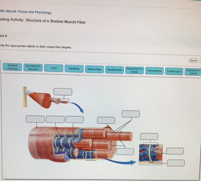
Art-labeling Activity: The Structure of a Skeletal Muscle Fiber Start studying Art-labeling Activity: The Structure of a Skeletal Muscle Fiber. Learn vocabulary, terms, and more with flashcards, games, and other study tools. ... Write. Test. PLAY. Match. Created by. BabeRuthless0504. Terms in this set (2) Art-labeling Activity: The Structure of a Skeletal Muscle Fiber... Art-labeling Activity: The Structure ...
Post a Comment for "42 art-labeling activity: structure of muscle tissues"