43 label the microscope parts
EOF microbenotes.com › parts-of-a-microscopeParts of a microscope with functions and labeled diagram Apr 19, 2022 · Figure: Diagram of parts of a microscope. There are three structural parts of the microscope i.e. head, base, and arm. Head – This is also known as the body. It carries the optical parts in the upper part of the microscope. Base – It acts as microscopes support. It also carries microscopic illuminators.
rsscience.com › stereo-microscopeParts of Stereo Microscope (Dissecting microscope) – labeled ... Labeled part diagram of a stereo microscope Major structural parts of a stereo microscope. There are three major structural parts of a stereo microscope. The viewing Head includes the upper part of the microscope, which houses the most critical optical components, including the eyepiece, objective lens, and light source of the microscope.

Label the microscope parts
› 6-label-the-microscopeLabel the microscope — Science Learning Hub Jun 08, 2018 · All microscopes share features in common. In this interactive, you can label the different parts of a microscope. Use this with the Microscope parts activity to help students identify and label the main parts of a microscope and then describe their functions. Drag and drop the text labels onto the microscope diagram. If you want to redo an ... animal cell microscope picture - As Funny Vodcast Photographs As you can see in the above labeled plant cell diagram under light microscope there are 13 parts namely Cell membrane. Cell Structure And Function. ... Browse 50454 cells microscope stock photos and images available or search for cancer cells microscope or blood cells microscope to find more great stock photos and pictures.. The diagram is very ... 14 Basic Parts of a Camera Explained - PhotographyAxis Parts of a Camera 1. Aperture. Aperture is the opening in front of the camera. It will be present in the lens part. For, an interchangeable lens camera, you will have the option to change the lens. So, you will have more options with the Aperture. But, for a standard point and shoot or bridge camera, the lens is a fixed one. So, the options are ...
Label the microscope parts. Biological Safety Cabinet (BSC): Types and Working Mechanism There are four types (A1, A2, B1, and B2 ) of Class II BSCs. The main differences between the types are the ratio of air exhausted from the BSC to the air that is recirculated within the BSC, and the type of exhaust system present. About 90% of all biosafety cabinets installed are Type A2 cabinets. There is a limited need for Class II Type B ... scheme work biology - Free KCPE Past Papers Draw and label the light microscope; Description of a cell; Drawing and labeling the light microscope . Light microscope; ... Golden tips Biology Page 15-16; KLB teachers book 1 pages 23-25 . 10. 1-2. THE CELL. Parts of the light microscope and their functions . Calculation of magnification using light microscope. By the end of the lesson, the ... 5 White Blood Cells Types and Their Functions - New Health Advisor 5. Basophils. Basophils are the least frequent type of white blood cell, with only 0-100 cells per mm 3 of blood. Basophils have large granules that perform functions that are not well known. They are very colorful when stained and looked at under the microscope, making them easy to identify. Chromosome - Genome.gov Definition. 00:00. 00:53. Chromosomes are threadlike structures made of protein and a single molecule of DNA that serve to carry the genomic information from cell to cell. In plants and animals (including humans), chromosomes reside in the nucleus of cells. Humans have 22 pairs of numbered chromosomes (autosomes) and one pair of sex chromosomes ...
Light Microscope (Assignment) - Amrita Vishwa Vidyapeetham The first experiment i.e.; knowing the parts of a microscope is a self explanatory animation. Instructor's can choose this animation in the class before detailing the parts of a microscope. To illustrate the working of a microscope, follow the instructions in the simulator. Students Assignment Parts of a Lab Report - Purdue University Parts of a Lab Report Note: Most 100-level chemistry labs require only worksheets to be filled out at the completion of each lab. Therefore, this information would be most useful for 200-level students as lab reports are often required for those courses. Now that you have completed an experiment and have collected all of the necessary ... 13 Best Image Annotation Tools of 2022 [Reviewed] - V7Labs LabelMe. Labelimg. VoTT. ImgLab. Every few months, a new training data platform enters the market promising new innovative features, faster labeling or higher accuracy. It's easy to get confused trying to choose the best image annotation tool for your specific use case. But—. Electron Microscope: Principle, Types, Applications - Microbe Online Electron microscope as the name suggests is a type of microscope that uses electrons instead of visible light to illuminate the object. Electromagnets function as lenses in the electron microscope, and the whole system operates in a vacuum. Since electrons have a very short wavelength, the resolving power of electron microscopes is very high ...
Light Microscope (Theory) - Amrita Vishwa Vidyapeetham Microscope can be separated into optical theory microscopes (Light microscope), electron microscopes (eg.TEM, SEM) and scanning probe microscopes. (eg.AFM, PSTM). Optical microscopes function on the basis of optical theory of lenses by which it can magnifies the image obtained by the movement of a wave through the sample. researchtweet.com › microscope-parts-labeledMicroscope, Microscope Parts, Labeled Diagram, and Functions Jan 19, 2022 · Microscope cell staining is a technique used to improve the visibility of cells and cell parts under a microscope. A nucleus or a cell wall can be seen more clearly by using different stains. 2. Iodine, crystal violet, and methylene blue are examples of simple stains. 3. Make a wet or dry mount with a coverslip. 4. virtuallabs.nmsu.edu › microVirtual Labs Bacteria Sampling (formerly Disposable Lab Equipment) should be followed by Gram Staining, then Using a Microscope Using the Microscope video play-through This work was supported by USDA CSREES and USDA National Institute of Food and Agriculture under two Higher Education Challenge Grant projects: 2008-38411-19055 and 2011-38411-30625. › wp-content › uploadsParts of a Microscope Printables - Homeschool Creations Parts of a eyepiece arm stageclips nosepiece focusing knobs illuminator stage objective lenses head base Label the parts of the microscope. You can use the word bank below to fill in the blanks or cut and paste the words at the bottom. Microscope Created by Jolanthe @ HomeschoolCreations.net eyepiece head objective lenses arm focusing knob base ...

Blood smear. Causes, symptoms, treatment Blood smear
› game › microscope-labelingMicroscope Labeling Game - PurposeGames.com This is an online quiz called Microscope Labeling Game. There is a printable worksheet available for download here so you can take the quiz with pen and paper. This quiz has tags. Click on the tags below to find other quizzes on the same subject.
![HLS [ Cartilage and Bone and Bone Histogenesis, compact bone] LOW MAG ...](http://www.bu.edu/histology/i/02701loa.jpg)
HLS [ Cartilage and Bone and Bone Histogenesis, compact bone] LOW MAG ...
Blood Cell Basics - Activity - TeachEngineering Discuss the various parts of the blood cell. Label these different parts on your drawing. Encourage the students to find these parts on their own drawings. Optional: Have students complete the Blood Cells Under a Microscope Vocabulary Worksheet. Assessment Pre-Activity Assessment
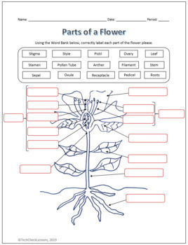
Parts of a Flower Labeling Science Worksheet for Google Slides | TpT
Metaphase - Genome.gov 00:00. 00:43. Metaphase is a stage during the process of cell division (mitosis or meiosis). Normally, individual chromosomes are spread out in the cell nucleus. During metaphase, the nucleus dissolves and the cell's chromosomes condense and move together, aligning in the center of the dividing cell. At this stage, the chromosomes are ...
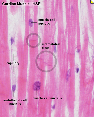
HM Practical - Cardiac Histology - Embryology
ECLIPSE Ti2 Series | Inverted Microscopes | Products | Nikon ... The ECLIPSE Ti2 inverted microscope delivers an unparalleled 25mm field of view (FOV) that revolutionizes the way you see. With this incredible FOV, the Ti2 maximizes the sensor area of large-format CMOS cameras without making compromises, and significantly improves data throughput. The Ti2's exceptionally stable, drift-free platform is ...
Post a Comment for "43 label the microscope parts"