39 label the internal features of stomach and duodenum using the hints if provided.
Medical Definitions - IFFGD Colonoscopy is a fiberoptic (endoscopic) procedure in which a thin, flexible, lighted viewing tube (a colonoscope) is threaded up through the rectum for the purpose of inspecting the entire colon and rectum and, if there is an abnormality, taking a tissue sample of it (biopsy) for examination under a microscope, or removing it. Colostomy Digestive lab Flashcards | Quizlet Label the mucous membrane tissue from the stomach using the hints if provided. Label the digestive abdominal contents using the hints if provided. Place the appropriate words and descriptions with the picture with the correct highlighted digestive accessory organ. Label the structures of the posterior thoracic wall using the hints if provided.
23.3 The Mouth, Pharynx, and Esophagus - Anatomy & Physiology In this section, you will examine the anatomy and functions of the three main organs of the upper alimentary canal—the mouth, pharynx, and esophagus—as well as three associated accessory organs—the tongue, salivary glands, and teeth. The Mouth The cheeks, tongue, and palate frame the mouth, which is also called the oral cavity (or buccal cavity).
Label the internal features of stomach and duodenum using the hints if provided.
25.1 Internal and External Anatomy of the Kidney On the superior aspect of each kidney is an adrenal gland. Each kidney looks like the kidney bean and the renal hilum is the entry and exit site for structures servicing the kidneys: vessels, nerves, lymphatics, and ureters. The medial-facing hila are tucked into the convex indentation of the kidney. Figure 25.1.2 Left Kidney. Internal Anatomy portal triad contents - emmeciquattro.net A portal triad in the liver consists of which three basic structures: a) a common bile duct, and branches of the right… Hepatic blood supply - Veterinary Histology Label the internal features of stomach and duodenum using the hints if provided. TAMU Biol 320: Module 9 Flashcards | Quizlet Image: Label the internal features of stomach and duodenum using the hints if provided.
Label the internal features of stomach and duodenum using the hints if provided.. Upper GI | Esophagram | Barium Swallow - Radiologyinfo.org Upper gastrointestinal tract radiography, also called an upper GI, is an x-ray examination of the esophagus, stomach and first part of the small intestine (also known as the duodenum). Images are produced using a special form of x-ray called fluoroscopy and an orally ingested contrast material such as barium. Pancreas histology: Exocrine & endocrine parts, function - Kenhub The pancreas is a large, mixed gland composed of five parts: the head, uncinate process, neck, body and tail. The location of the pancreas is mostly retroperitoneal, except for the tail. This organ extends from the C-shaped curve of the duodenum, passes behind the stomach and finishes at the hilum of the spleen. Digestive System Flashcards | Quizlet Label the non-digestive abdominal contents using the hints if provided. ... Label the internal features of stomach and duodenum using the hints if provided. DIGESTIVE Flashcards | Quizlet Label the structures and regions of the small intestine. Image: Label the structures and ... Label the stomach and duodenum using the hints if provided.
GI Post Lab Flashcards | Quizlet Label the internal features of stomach and duodenum using the hints if provided. Label the histologic features of the duodenum using the hints if provided. Label the light micrograph of the colon using the hints provided. Label the light micrograph of the liver using the hints provided. ReaderUi ReaderUi Module 3 Study Guide ch 24 ch 25Catrina Greene BIO-169 ... - Course Hero Label the parts of the liver and gallbladder using the hints provided. Label the abdominal contents using the hints provided. Label the sagittal section of the mesenteries. Label the mucous membrane tissue from the stomach using the hints if provided. Correctly organize the events of the defecation reflex. 007460.pdf - LESSON 5 THE HUMAN BODY In the fifth lesson ... - Course Hero Ingestion, digestion, absorption, and elimination are the four phases of digestion. Iningestion, you literally put the food into your digestive system when it enters your mouth. Indigestion, food is broken down mechanically by chewing and chemically by juices that target the breakdown of certain substances.
Digestive lab Flashcards | Quizlet Label the mucous membrane tissue from the stomach using the hints if provided. Label the digestive abdominal contents using the hints if provided. Place the appropriate words and descriptions with the picture with the correct highlighted digestive accessory organ. Label the structures of the posterior thoracic wall using the hints if provided. The Dogfish Shark—Structure and FUNction! - Carolina.com The stomach's longitudinal folds, called rugae, allow the stomach to expand. Discuss these digestive structures in light of the fact that the shark does not chew its food but instead bites off and swallows large chunks of it. At a J-shaped turn along the digestive tube, the stomach leads into the duodenum. TAMU Biol 320: Module 9 Flashcards | Quizlet Label the internal features of stomach and duodenum using the hints if provided. Image: Label the internal features of stomach and duodenum using the hints ... portal triad contents - rfclark.com Automation Products | Pittsburgh, PA | Russel F. Clark; coiled usb-c cable with aviator; how to keep a german shepherd in your yard; who is a better medical ninja sakura or tsunade
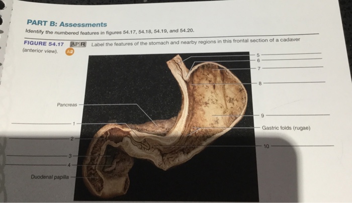
Solved: PART B: Assessments Identify The Numbered Features... | Chegg.com
Practical 2 Flashcards | Quizlet label the internal features of stomach and duodenum using the hints if provided. Image: label the internal features of stomach and duodenum using the hints ...
Post a Comment for "39 label the internal features of stomach and duodenum using the hints if provided."